Introduction
Stroke is a clinical syndrome of vascular origin characterized by the sudden onset of focal neurological signs.1 It represents a significant public health issue globally, as it is one of the leading causes of death and disability.2 Stroke survivors often experience a decline in their quality of life due to various sequelae, with depression being the most common neuropsychiatric consequence.3
Post-stroke depression is diagnosed in approximately more than one-third of all stroke survivors,4,5 and it is considered the main predictor of poor functional outcome.3 Among the multiple factors underlying post-stroke depression, several studies have explored the relationship between the location of the stroke lesion and the development of this sequelae. Their results suggest that stroke lesions in the left cerebral hemisphere as well as in frontal regions lead to depression.3,6 To study post-stroke depression in animal models, the left medial prefrontal cortex has also been suggested as a region of interest.7
Even though the middle cerebral artery occlusion model in combination with chronic mild stress is considered a valid pre-clinical model for the study of post-stroke depression,8 it has been observed that this model can produce motor deficits that interfere with the evaluation of depression-like behavior.7 Taking that into consideration, this study hypothesized that the photothrombotic lesion technique could be a useful model for the study of post-stroke depression. Knowing that it facilitates the behavioral study of ischemic stroke consequences in specific anatomical-functional regions9 since it allows inducing focal lesions in the cortical10 or subcortical11 brain area of interest.
To induce a stroke, this technique involves administering a photosensitive dye (such as Rose Bengal), followed by illuminating the brain through the skull. The dye, activated by the light, generates singlet oxygen and superoxide. This results in endothelial injury and platelet activation and aggregation, ultimately leading to ischemic cell death and the formation of a cortical ischemic lesion.11
Methods
An experimental study using independent groups was conducted between August and December 2023. The animals were treated in accordance with the specifications for the production, care, and use of laboratory animals (NOM-062-ZOO-1999), and the research protocol approved by the Internal Committee for the Care and Use of Laboratory Animals of the National Institute of Neurology and Neurosurgery Manuel Velasco Suárez (88/18) was followed.
The sample size was determined based on previously published studies employing similar methodologies to ensure methodological consistency and comparability of results.12,13 This study adhered to the ARRIVE guidelines to enhance transparency and reproducibility in animal research. All efforts were made to minimize the number of animals used while maintaining statistical rigor and ensuring reliable outcomes. In terms of study design, a negative control was implemented by comparing sham surgery with stroke surgery, enabling the identification of differences attributable to the photothrombotic lesion. To maintain data integrity, rats that developed otitis post-surgery were excluded based on predefined exclusion criteria. Randomization was conducted using a computer-based random order generator to assign each rat to either the sham or stroke group.
A timeline of the experimental design is provided in figure 1, summarizing the key steps: habituation, baseline, and evaluation phases for the forced swim test (FST), as well as the timing for the sucrose preference test (SPT) and beam walking test (BWT). This figure aims to facilitate understanding of the protocol and clarify the methodological approach.
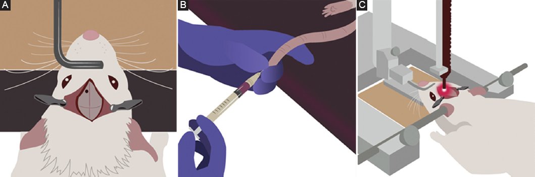
Figure 1. Photothrombotic lesion procedure. A: rats were anesthetized with a combination of ketamine (90 mg/kg) and xylazine (8.5 mg/kg) prior to stereotaxic surgery. A trephine was made on the skull to target the left medial prefrontal cortex. B: rose Bengal (or saline solution for the sham group) was administered intravenously. C: a cold light source was placed over the trephine for 20 min.
Forty-three male Wistar rats (weighing 220-390 g) were used in this study. The rats were randomly divided into two groups: the sham group (n = 20) and the stroke group (n = 23). They were primarily pair-housed in standard acrylic cages under a 12-h light-dark cycle, with ad libitum access to food and water. However, during the SPT, they were temporarily housed individually.
Rats were anesthetized with a combination of ketamine (90 mg/kg) and xylazine (8.5 mg/kg) prior to stereotaxic surgery. A trephine was made on the skull to target the left medial prefrontal cortex at the following coordinates in reference to the bregma:14 AP = +3.2, LM = +1.6. Subsequently, a 0.5 mL/kg dosage of a previously prepared 15 mg/mL Rose Bengal and saline solution was administered intravenously to the stroke group. A 0.5 mL/kg dose of saline solution was used for the sham group. The administration was allowed to diffuse into the bloodstream for 5 min.15,16 A fiber-optic bundle of a cold light source (KL 1500 LCD, Schott, Germany) with a 3 mm aperture was placed over the trephine for 20 min (Fig. 2). Post-surgery, a hemostatic sponge was placed at the trephine site, and the wound was sutured. The rats were given a subcutaneous dose of tramadol and saline solution for pain management. The animals’ temperature was regulated throughout all the procedures using a thermal pad.
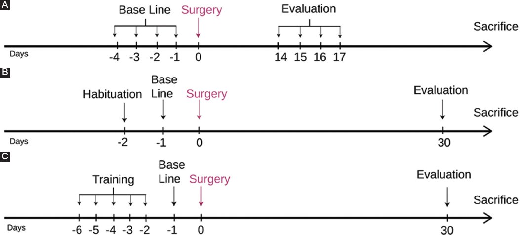
Figure 2. Experimental timeline: (A) Arrows indicate days on which the sucrose preference test, (B) forced swim test, (C) and beam walking test were conducted.
Depression-like behavior was evaluated at 14 days post-stroke (14 DPS) using a SPT protocol adapted from the one established by De la Tremblaye et al.,17 and at 30 DPS (30 DPS) using the FST protocol described by Yankelevitch-Yahav et al.18
Given the high prevalence of motor deficits after an ischemic stroke,19 the BWT was conducted to minimize potential biases while evaluating FST performance. Rats were trained to cross the beam for 5 days prior to establishing the baseline. Motor performance was assessed at 30 DPS using the scores described by Ramos-Languren et al. (Fig. 2).20
IBM Statistical Package for the Social Sciences Statistics ver. 29.0 (IBM Co., Armonk, NY, USA) was used for statistical analysis. The results obtained in the behavioral tests were analyzed with the Mann-Whitney U test and expressed as the mean ± standard error of the mean. In all cases, the level of significance was set at p < 0.05.
Results
Of the 45 Wistar rats initially enrolled in the study, two died prior to group allocation due to unrelated complications, leaving a final sample of 43 animals divided into two groups: 20 sham and 23 stroke. Over the course of the experimental protocol, an additional five rats (two from the sham group and three from the stroke group) were lost due to post-surgical complications or humane euthanasia criteria. Consequently, the final sample size for statistical analyses comprised 18 stroke and 17 sham rats, reflecting survival rates of 78.26% and 85%, respectively.
Mortality and survival analysis
The mortality rates observed in both groups deviated from previous studies using similar methodologies, where 100% survival was reported. The increased mortality in the sham group (15%) and the stroke group (21.7%) highlights potential complications related to surgical stress or individual variability rather than the lesion itself (Fig. 3).
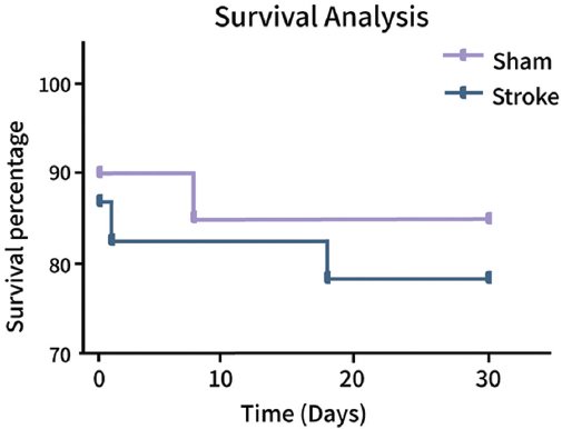
Figure 3. Percentage of survival along the experimental procedure. Comparison between sham and stroke groups.
Behavioral tests
SPT
At 14 days post-surgery, stroke rats exhibited a significant reduction in sucrose consumption compared to sham rats (U = 90, p = 0.023), indicating anhedonia-like behavior. Stroke rats consumed an average of 67.3 ± 5.1% sucrose solution, whereas sham rats consumed 81.4 ± 3.8% (Fig. 4). This result aligns with previous findings suggesting an early onset of depression-like behaviors in photothrombotic models.
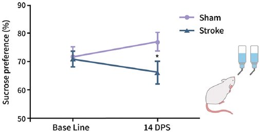
Figure 4. Sucrose preference test analysis. It was observed that there was a decrease in sucrose preference in photothrombotic lesioned rats, suggesting anhedonia. Mann-Whitney’s U (*p = 0.023).
FST
Immobility times were measured at 30 DPS to evaluate depression-like behavior. However, no significant differences were observed between the groups (U = 107, p = 0.289). Stroke rats displayed an average immobility duration of 168.7 ± 12.5 s, compared to 160.3 ± 10.2 s in sham rats. This lack of significance suggests that the depressive-like phenotype observed in the SPT may not be robustly reflected in this behavioral test, potentially due to methodological factors or the timing of evaluation (Fig. 5).
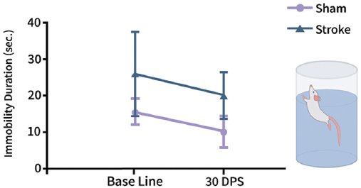
Figure 5. Forced swim test. No significant differences were observed in immobility duration, suggesting no changes in motor performance.
BWT
To rule out motor impairments as a confounding factor for the FST, motor performance was assessed at 30 days post-surgery. No significant differences in beam traversal times were found between groups (U = 130, p = 0.825). Stroke rats required an average of 7.3 ± 0.6 s to traverse the beam, whereas sham rats took 7.1 ± 0.5 s (Fig. 6). This result supports the hypothesis that the photothrombotic lesion did not induce substantial motor deficits, allowing a more targeted assessment of depression-like behaviors.
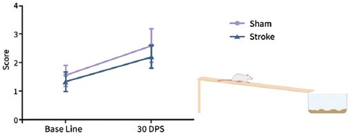
Figure 6. Beam walking test. No significant differences were found in beam walking test scores, showing the absence of motor effects of the photothrombotic lesion.
Discussion
Regarding the need to develop new pre-clinical models for post-stroke depression research, this study tested the usefulness of the photothrombotic lesion technique. To evaluate whether an ischemic stroke induced by photothrombosis promoted the development of a depression-like phenotype in rats, the SPT and the FST were used. In addition, the BWT was employed to rule out any potential biases in the FST results due to post-stroke motor alterations.
Although one of the major drawbacks of the photothrombotic technique is the limited or absent ischemic penumbra, this model was selected for its significant advantages, such as the ability to target a specific cortical brain region for the ischemic lesion, its simplicity of execution, minimally invasive nature, and low mortality rate.21
Regarding the survival rate, while previous studies have reported a 100% survival rate for the animals using this technique,22 our study observed a lower survival rate (78.26%) in stroke-induced rats. However, it is important to highlight that the strokes were induced in different brain regions, which may account for the variation. Therefore, in this study, the higher mortality observed in the sham group (15%) compared to previous reports may be attributed to surgical stress or individual variability. Importantly, all post-surgical care, housing conditions, and testing protocols were identical for both groups, reducing the likelihood of methodological bias.
As for the behavioral assessment, the differences found in the SPT between groups 2 weeks after surgery are consistent with previously reported findings.17,23 Conversely, the results obtained in the FST differ from previous studies, which showed an increased immobility time between weeks 3 and 5 post-stroke.23,24
Post-stroke depression is not only the most common neuropsychiatric sequel but also a major factor in poor functional and cognitive recovery in survivors.3 Therefore, it is crucial to conduct research that enhances our understanding of this condition to reduce the sequelae and complications for the patients25 and develop new and more specific treatments.26 Although still under development,27 pre-clinical models play a significant role in achieving these goals.4,25,28
Rodents are often the primary choice for animal models in cerebrovascular disease research;10,23 however, it is crucial to recognize that they are not merely miniature versions of humans.29 This distinction is significant because stroke lesions in humans are confined to the white matter, whereas rodent studies on white matter are limited due to its reduced volume.30 Moreover, within the study of depression, some subjective and complex aspects experienced by humans, such as low self-esteem or suicidal ideation, cannot be modeled in rodents.29
Regarding the behavioral assessment, it is important to note that although the SPT is considered the best method to evaluate anhedonia in rodents, its lack of standardized equipment makes it difficult to control water loss, potentially affecting experimental results.31 Conversely, repeated exposure to the FST can lead to inaccurate results.29 In this project, animals underwent the test on three occasions to adhere to the established protocol, which may explain the lack of significant differences between groups.
Although the photothrombotic lesion technique allows for the induction of a focal stroke, no technique was employed to evaluate the volume of the infarction. Given that the peri-infarct cortex is connected to other brain regions,11 Nissl staining could be a valuable tool for enhancing future studies. It is also essential to consider that while many studies associate left hemisphere lesions with post-stroke depression,3 there are also reports linking right hemisphere lesions to PSD.6,7 Therefore, it may be important to induce lesions in the right hemisphere and compare the resulting behavioral outcomes with those observed in this study. Furthermore, future research should evaluate the similarities and differences between male and female rodents, as well as among young and old rodents and different strains.28 This approach will help determine whether these factors influence the characteristics of infarction and/or the development of post-stroke depression. Since motor deficits in the early days post-lesion were not assessed in this study, future research could incorporate longitudinal motor evaluations to provide a more comprehensive understanding of functional recovery.
Regarding the behavioral assessment, it would be beneficial to evaluate depression-like behavior using the SPT at multiple time points to ascertain its onset and duration. Previous studies have demonstrated a decreased sucrose preference 1 week32–34 and 28 days34,35 after implementing the post-stroke depression model.
Given the inconclusive results from behavioral tests, it is recommended that future studies evaluate additional parameters associated with depression-like behavior following a stroke. These include body weight,17,35 food intake,17 and various depression biomarkers such as inflammatory cytokines,36 corticosterone levels,17 hypothalamic-pituitary-adrenal axis hormones,37 and brain-derived neurotrophic factor levels.27,33,35,36,38
Several limitations of this study must be acknowledged. The higher mortality observed in the sham group raises concerns about potential surgical stress or individual variability. In addition, the lack of early motor assessments limits the understanding of initial recovery patterns. Although the photothrombotic lesion technique offers precise targeting, it presents challenges such as a narrow ischemic penumbra and variability in infarct size, which may affect its translational value. Another limitation is the lack of a formal sample size calculation based on Festing’s criteria. While the sample size was selected based on previously published studies with similar methodologies to ensure comparability and methodological consistency, future research should incorporate a priori power analyses to optimize the number of animals used while maintaining statistical robustness and ethical considerations. Standard behavioral tests, such as the SPT and FST, may not fully capture the complexity of depression-like behavior, highlighting the need for complementary biochemical and histological evaluations. Addressing these limitations in future research will enhance the utility and validity of this pre-clinical model.
Conclusions
The high incidence of post-stroke depression highlights the need to conduct research aimed at mitigating its impact on survivors’ quality of life. Although pre-clinical studies are essential for this purpose, the limitations of current models make it crucial to explore new alternatives. This study proposed that the photothrombotic lesion technique could be a promising method for investigating post-stroke depression due to its various advantages. However, because the results from behavioral tests were not sufficiently conclusive, it is recommended that future research incorporate additional complementary techniques, such as depression biomarkers, to better determine the presence of a depression-like behavioral phenotype following a photothrombotic stroke.
Funding
This research was funded by a grant from the General Directorate of Academic Personnel Affairs (DGAPA) of the National Autonomous University of Mexico (UNAM), through the Support Program for Research and Technological Innovation Projects (PAPIIT)-IN216221.
Conflicts of interest
The authors declare no conflicts of interest.
Ethical considerations
Protection of human subjects and animals. The authors declare that the procedures followed were in accordance with the ethical standards of the responsible committee on human experimentation and with the World Medical Association and the Declaration of Helsinki. The procedures were authorized by the Institutional Ethics Committee.
Confidentiality, informed consent, and ethical approval. This study does not involve personal patient data, medical records, or biological samples, and does not require ethical approval. SAGER guidelines do not apply.
Declaration on the use of artificial intelligence. The authors declare that no generative artificial intelligence was used in the writing or creation of the content of this manuscript
