

Introduction
The tendon is a specialized form of connective tissue and an integral part of the musculoskeletal system, connecting muscle to bone. This tissue, which is sensitive to mechanical stimuli, possesses distinct mechanical characteristics that enable it to react and adjust to the forces exerted by muscle.1 They can resist the tensile solid forces carried on by movement and preserve elastic energy.2 Tendon disorders vary from long-term injuries to sudden, acute injuries and can include partial or complete ruptures of the tendon.3
Peritendinous tissue adhesion following tendon injury is a significant complication that causes pain as well as movement dysfunction, often requiring complex surgical intervention.4 It is still extremely challenging to successfully heal tendons without adhesions, even with improvements in surgical methods and post-operative rehabilitation methods. Post-operative problems such as tendon rupture and loss of motion continue to be an issue for both patients and surgeons.5 Numerous rehabilitation protocols and surgical repair techniques have been attempted to manage these complications.6 In addition, various synthetic materials, which include silicone, polyethylene membranes, alumina barriers, polytetrafluoroethylene, and polyhydroxy-ethyl methyl-methacrylate membranes, have been utilized to prevent the fibrosis formation that results in adhesion. However, none of these have been successful enough for clinical use.7–9
Lactic acid and glycolic acid monomers are copolymerized to form poly(lactic-co-glycolic acid) (PLGA), an aliphatic polymer with a polyester backbone. As a copolymer, PLGA can be synthesized into various formulations, taking into account the lactic and glycolic acid ratio in its conformation.10 Polycaprolactone (PCL), which is made up of repeated hexanoate units, is another polymer in the polyester class. PCL is produced through either polymerization of ε-caprolactone with Sn(Oct)2 as a catalyst or polycondensation of 6-hydroxyhexanoic acid.11 Today, various applications of these polymers, such as in drug and vaccine delivery, have been explored.12–14 Methylprednisolone (MTP) is a synthetic glucocorticoid used in the treatment of disorders associated with fibrosis, including hypertrophic scars and keloids, due to its antifibrotic effects.15 However, they are not so innocent drugs. Corticosteroids are well-known for disrupting the wound-healing process and causing several systemic side effects such as diabetes, hypertension, gastritis, and cataracts.16
In this study, the MP-loaded PLGA formed the outer layer of the barrier, which was aimed at reducing the peritendinous adhesion. The PCL layer (inner surface) was designed to be on the tendon body to prevent the transmission of MP toward the tendon surface. The impact of this design on tendon recovery and the development of adhesions was examined.
Methods
Experimental method for the production of MP-loaded PLGA/PCL nanofibers
Local Ethics Committee approval was obtained for this study (protocol no: 2023-028). This study was conducted in accordance with the ethical principles of the Declaration of Helsinki. In our previous study, it was determined that the optimal dose loaded into the PLGA mat was 25% of the PLGA weight (g). Lower doses were linked to insufficient effects, while higher doses resulted in the atrophic effects of MP.17 The electrospinning procedure was carried out for both PCL and 25%MP-PLGA layers of the adhesion barrier mat.
Physical characteristics of electrospun nanofibers
At least 100 random fibers were analyzed on scanning electron microscopy (SEM) images in order to determine the average diameter of each sample. A Fourier transform infrared spectroscopy (FT-IR) spectrometer was utilized to identify characteristic functional groups of both nanofiber layers in the range of 400-4000 cm−1. The hydrophilicity characteristics of the nanofibers were assessed by a device of water contact angle meter (Biolin Scientific Attention, Theta Lite) by dripping 5 μL of deionized water into each nanofiber sample. The tension test was conducted using a texture analyzer to evaluate the mechanical properties of the samples in a size of 10 × 50 mm2 and 6 mm/min tensile velocity in accordance with ASTM D638-14 standard.
In vitro biodegradability analysis
The biodegradability analysis involved evaluating pure PCL nanofibers, 25% MP-PLGA, and 25% MP-PLGA/PCL nanofiber mats in a hydrolytic environment made of polybutylene succinate (PBS) (pH 7.4) over a period of 8 weeks. Each nanofiber mat sample, measuring 30 × 30 mm, was immersed in 15 mL of PBS and incubated in tubes at 37°C. The residual medium was replaced every week, and mass changes in the related nanofibers were tracked through gravimetric measurements.
In vitro drug release behavior
The drug release characteristics of samples containing 25% MP-PLGA and 25% MP-PLGA/PCL were investigated by submerging them in PBS, as detailed in a prior study.17
The investigation focused on the drug release kinetics of 25% MP-PLGA and 25% MP-PLGA/PCL, employing five distinct mathematical models. The analysis demonstrated that the initial 60% of experimental release data adhered to the Korsmeyer-Peppas model.18 The optimal model was ascertained through the comparison of the fit correlation coefficient (R2) of each model with the observed data.
In vivo study of Achilles tendon repair
PREPARATION OF THE TENDON INJURY
Forty-two Wistar albino rats, aged 4 months, were classified into four groups, with 14 Achilles tendons allocated to each group. In Group 1 (sham group), the right Achilles tendons were left untreated, with no intervention applied. In Group 2 (control group), the left tendons were transected and then repaired. The surgical repair involved the modified Kessler technique and primary saturation using 5-0 Polypropylene sutures. In Group 3, the left tendons were repaired using the same technique and then wrapped with neat PLA/PLGA. In Group 4, the left tendons were repaired and then wrapped with 25% MP-PLGA/PCL.
At the end of 4 weeks after surgery, the tendons were retrieved by accessing the old incision. A primary macroscopic visual assessment (Table 1) of adhesions was conducted. Each group was subsequently split into two: an equal number of tendons (7 for each) were allocated for biomechanical and histopathological examination. The atrophic effects of the methylprednisolone were evaluated by measuring tendon length. The systemic effects of methylprednisolone were assessed by measuring glucose levels at three points: before surgery, 2 weeks after surgery, and prior to sacrifice.
Table 1. Criteria described by Tang et al., for microscopic and macroscopic evaluation of adhesions
| Points | Criteria for macroscopic evaluation |
|---|---|
| Features of adhesion | |
| Quantity (length) 0 1 2 3 | No apparent adhesionsLocalized, longitudinal extension within 10 mmLongitudinal extension between 10 and 15 mmExtensive, longitudinal extension beyond 15 mm |
| Quality (characteristic) 0 1 2 3 | No apparent adhesionsLoose, elastic, and largely movableModerately, dense, and movableDense, rigid, and not movable |
| Grade of adhesions (severity) 0 2 3,4 5,6 | NoneSlightModerateSevere |
| Criteria for histopathological evaluation | |
| Points | Histological appearance |
| Inflammation intensity 0 1 2 3 | NoneMildModerateSevere |
| Vascularization: ×1 magnification 1 (mild) 2 (moderate) 3 (severe) | 0–5 pieces capillaries6-10 pieces capillariesMore than 10 capillaries |
| Fibrosis 0 1 2 3 | NoneMildModerateSevere |
| Mucin accumulation 0 1 | NoYes |
HISTOPATHOLOGICAL EVALUATION
Samples were stained with hematoxylin and eosin (H&E), Masson trichrome (MTK), and Alcian blue. The slides prepared for histochemical staining were incubated overnight at 50-55°C. The slides were examined under a light microscope (Table 1). In the H&E-stained slides, the density and type of inflammatory cells, as well as vascularization, were assessed; fibrosis was evaluated in the MTK-stained slides, and mucin accumulation in the Alcian blue-stained slides.19 The tendon healing score was given based on the presence of granulation tissue and collagen alignment; a score of 1 indicates excellent healing while a score of 3 indicates poor healing.
BIOMECHANICAL EVALUATION
Seven tendons from each group were allocated for this analysis. The tests were performed with a tensile analysis device with a 1 mm/min pulling speed. The tensile properties of the tendons, including maximum load and breaking load, were analyzed.
Statistical analysis
The data were analyzed using the Statistical Package for the Social Sciences 24.0 software. A normality test was conducted. For group comparisons, the Mann-Whitney U test and Wilcoxon signed-rank test were applied. Kruskal-Wallis variance analysis was used for comparisons among multiple groups. In post hoc pairwise comparisons, the Mann–Whitney U test with Bonferroni correction was used. A p-value below 0.05 was considered statistically significant for all analyses.
Results
Physiochemical and morphological evaluation of 25% MP-PLGA/PCL nanofibers
SEM ANALYSIS
SEM images of both nanofiber layers of PCL magnified at 10k and 50k and 25% MP-PLGA, magnified at 2k and 10k, are shown in figure 1. The SEM analysis revealed that the electrospinning process produced uniform, bead-free, and smooth drug-loaded PLGA and PCL nanofiber membranes as layers. The PCL nanofibers had a relatively narrow mean diameter of 187.1 ± 39 nm, whereas the 25% MP-PLGA nanofibers exhibited a broader diameter, measuring 550.5 ± 105 nm.
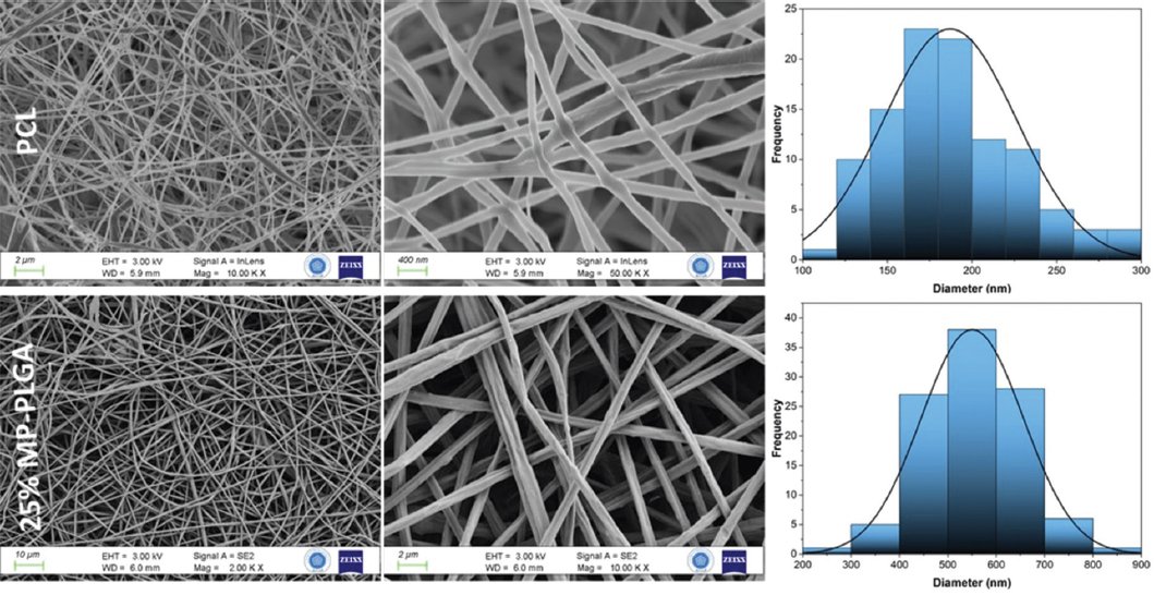
Figure 1. Scanning electron microscopy images of polycaprolactone and 25% MP-poly(lactic-co-glycolic acid) nanofibers with their diameter distribution curves.
FTIR ANALYSIS
The FTIR analysis, as shown in figure 2, revealed that 25% MP-PLGA exhibited an ester carbonyl stretch (C=O) with a prominent peak at 1749 cm-1, indicating no significant shift due to MP interaction. Additional major peaks were observed around 1452 cm-1 and 1382 cm-1, corresponding to C-H bending vibrations, along with peaks at 1176 cm-1 and 1086 cm-1, which were identified as asymmetrical C-O stretching vibrations characteristic of PLGA.20,21 Absorption peaks at 2996 cm-1 and 2937 cm-1 were also noted, corresponding to the aliphatic C-H stretching vibrations of methyl groups. The peaks at 2944 cm-1 and 2867 cm-1 in the PCL spectrum are attributed to the stretching vibrations of C-H bonds.22 Peaks at 1723 cmv, 1295 cm-1, and 1243 cm-1 were due to carbonyl stretching, C-O and C-C stretching, and asymmetric C-O-C stretching, respectively. In addition, peaks at 1184 cm-1 and 1164 cm-1 were related to C-O-C stretching.23
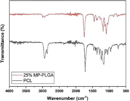
Figure 2. Fourier transform infrared spectroscopy spectrum of polycaprolactone and 25% MP-poly(lactic-co-glycolic acid) nanofibers.
MECHANICAL TEST
The stress-strain mechanical curves of pure PCL, 25% MP-PLGA, and 25% MP-PLGA/PCL nanofibers were evaluated. The maximum stresses (N/mm2) observed were 6.6, 0.95, and 2.56 MPa, corresponding to maximum strain (%) values of 166.3%, 17.97%, and 96.4%, and Young’s modulus values of 5.07 MPa, 10 MPa, and 3.2 MPa for neat PCL, 25% MP-PLGA, and 25% MP-PLGA/PCL nanofibers, respectively. The final product, 25% MP-PLGA/PCL nanofibers, exhibited tensile strength between that of PCL and 25% MP-PLGA, with improved ductility of 96.4% due to the inclusion of PCL, compared to 25% MP-PLGA.
Hydrophilicity behavior analysis regarding water contact angle measurement, in vitro drug release study, and biodegradability assessment
Hydrophilicity measurements for both sides of the PCL-25% MP-PLGA nanocomposite scaffold adhesion barrier model were performed. The water contact angles of PCL and 25% MP-PLGA nanofibers were recorded as 128.5° and 94.8°, respectively, as depicted in figure 3A.
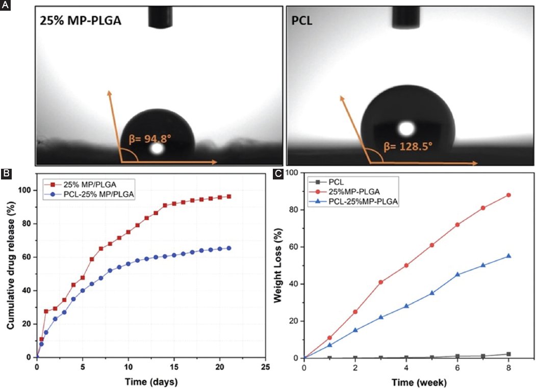
Figure 3. A: WCA of PCL and 25% MP-PLGA nanofibers. B: biodegradability characteristics of PCL, 25% MP-PLGA, and 25% MP-PLGA/PCL nanofibers. C: in vitro drug release of 25% MP-PLGA and 25% MP-PLGA/PCL nanofibers. PLGA: poly(lactic-co-glycolic acid); PCL: polycaprolactone.
The drug release behavior of 25% MP-PLGA and 25% MP-PLGA/PCL nanofiber samples over a 21-day period is presented in figure 3B. Within the first 24 h, the 25% MP-PLGA sample demonstrated a more pronounced initial burst effect compared to the PCL-blended sample. Cumulative drug release analysis during the first 24 h indicated release rates of 27.6% for the 25% MP-PLGA sample and 15% for the 25% MP-PLGA/PCL sample. By the end of the 21-day period, 96.3% of the drug had been released from the 25% MP-PLGA sample, whereas 65.4% was released from the 25% MP-PLGA/PCL sample.
The drug release mechanism of both the 25% MP-PLGA and 25% MP-PLGA/PCL nanofibers was evaluated according to different kinetic models. Regression coefficients (R2) derived from these models representing in vitro drug release data were presented in table 2.
Table 2. R2 values calculated from kinetic models of MP release data of MP-loaded samples
| Samples | Zero-order | First-order | Higuchi | Korsmeyer-Peppas | Hixson-crowell |
|---|---|---|---|---|---|
| 25% MP/PLGA | 0.8881 | 0.9896 | 0.9831 | 0.9121 | 0.9845 |
| 25% MP-PLGA/PCL | 0.8202 | 0.9027 | 0.9831 | 0.9905 | 0.8776 |
|
PLGA: poly (lactic-co-glycolic acid); PCL: polycaprolactone. |
|||||
To assess the biodegradability of the nanofibers, samples were immersed in PBS for a period of 8 weeks. When the degradation process was completed, the neat PCL, 25% MP-PLGA, and 25% MP-PLGA/PCL nanofiber mats showed weight losses of 2.2%, 88%, and 55%, respectively, compared to their initial weights, as shown in figure 3C.
In vivo assessment of 25 MP-PLGA/PCL nanofiber for tendon repair
BLOOD GLUCOSE LEVEL
The average pre-operative blood glucose levels for groups 2-4 were 91 ± 5.38, 88.5 ± 5.62, and 89 ± 6.24, respectively. In the 2nd week, these values were 90.35 ± 6.28, 90 ± 7.54, and 93.28 ± 8.2, respectively. Before sacrifice, the values were 92.5 ± 8.7, 89.42 ± 5.43, and 88.35 ± 6.11. No statistically significant variances were detected either within or between the groups (p > 0.05).
MACROSCOPIC ANALYSIS
The outcomes of the macroscopic examination are provided in table 3. When comparing the groups, a statistical variance was detected between Group 1 and all other groups regarding adhesion length, adhesion characteristics, and adhesion degree (p < 0.05). There was no statistically significant difference observed between Groups 2 and 3 in terms of these parameters (p > 0.05).
Table 3. Results of macroscopic and microscopic evaluations. There were 14 rats in each group for macroscopic evaluation and seven rats in each group for biomechanical and microscopic evaluation
| Parameter | Group 1 | Group 2 | Group 3 | Group 4 |
|---|---|---|---|---|
| Macroscopic evaluation Quantity (length) Quality (characteristic) Grade of adhesions (severity) Tendon length (MM) | 0005.67 ± 0.66 | 2.5 ± 0.762.64 ± 0.634.9 ± 1.265.46 ± 0.57 | 2.57 ± 0.752.64 ± 0.495.28 ± 1.265.39 ± 0.48 | 1.2 ± 0.801.5 ± 0.852.71 ± 1.855.64 ± 0.63 |
| Biomechanical evaluation Maximum force (N) Maximum pressure (N/MM2) | 53.8 ± 5.115.6 ± 0.90 | 48.2 ± 7.614.6 ± 0.75 | 39.1 ± 4.212.43 ± 1.14 | 7 ± 8.314.5 ± 0.88 |
| Microscopic evaluation Inflammation intensity Vascularization Fibrosis Mucin accumulation Tendon healing score | 01 ± 0.00001 ± 0.00 | 1.85 ± 0.372.71 ± 0.482.57 ± 0.530.71 ± 0.482.14 ± 0.69 | 2.57 ± 0.532.57 ± 0.532.85 ± 0.370.85 ± 0.372.71 ± 0.48 | 2.14 ± 0.692 ± 0.572.14 ± 0.370.71 ± 0.481.57 ± 0.53 |
A statistically significant difference was identified between Groups 2 and 4, as well as between Groups 3 and 4 (p = 0.007, 0.004, respectively) in terms of adhesion length. Adhesion characteristics (p = 0.009, 0.012, respectively) and the degree of adhesion (p = 0.021, 0.005, respectively). The values of tendon lengths did not exhibit any statistical difference (p > 0.05). A foreign body reaction characterized by abscess formation and mat exposure was observed in 12 out of 14 animals in Group 3 (85%). In some of these cases, it was observed that the neat PLGA/PCL mats had not dissolved (Fig. 4).
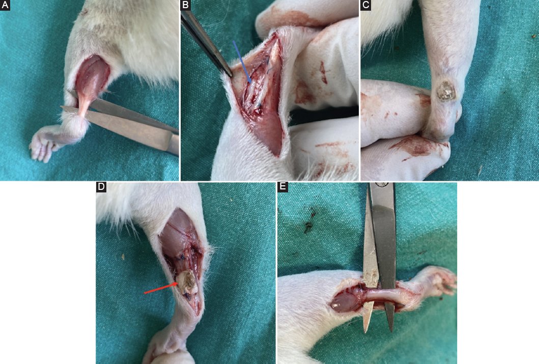
Figure 4. A: healthy tendon with no surgical intervention from Group 1. B: peritendinous adhesion (indicated by the blue arrow) observed in Group 2. C and D: abscess formation and persistent poly(lactic-co-glycolic acid)/polycaprolactone mat (indicated by the red arrow) observed in Group 3. E: smooth tendon healing observed in Group 4.
BIOMECHANICAL EVALUATION
Seven tendons from each group were tested using a texture analyzer. Values are presented in table 3. In terms of mean maximum breaking weight and tensile strength, there was no significant difference between the other groups (p > 0.05). However, there was a significant difference between Group 3 and all other groups (p < 0.05).
MICROSCOPIC ANALYSIS
Group 1 did not show any signs of inflammation, fibrosis, or mucin accumulation. The mean inflammatory cell densities for Groups 2-4 were 1.85 ± 0.37, 2.57 ± 0.53, and 2.14 ± 0.69, respectively (Figs. 5 and 6). A statistically significant difference was found between Group 1 and all other groups (p < 0.05) and between Groups 2 and 3 (p = 0.02).
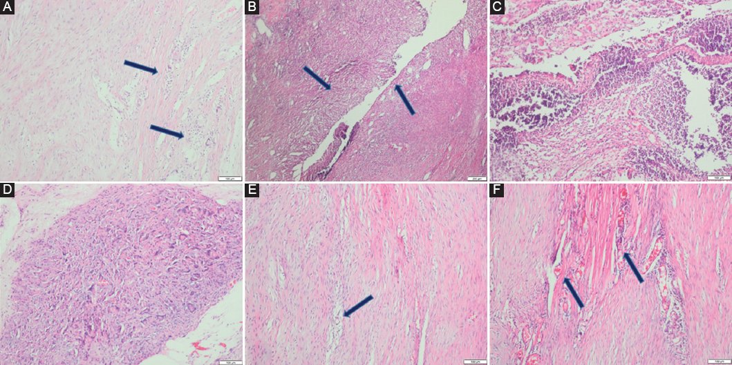
Figure 5. A: ×100 H&E: mild inflammation. B: ×40 H&E: severe inflammation. C: ×100 H&E: abscess. D: ×100 H&E: Foreign body-type inflammatory granulation tissue. E: ×100 H&E: mild vascularization. F: ×100 H&E: severe vascularization. H&E: hematoxylin and eosin.
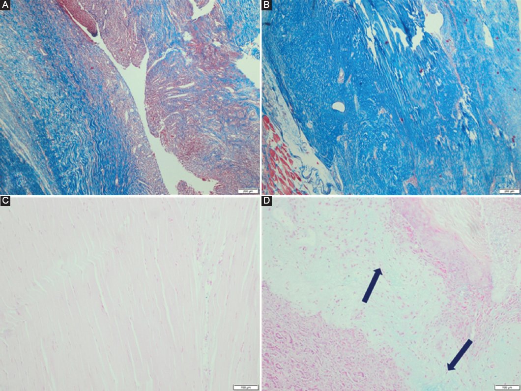
Figure 6. A: ×40 MTK: Moderate fibrosis. B: ×40 MTK: Severe fibrosis. C: ×100 Alcian blue: No mucin. D: f ×200 Alcian blue: positive mucin. MTK: Masson trichrome.
The mean vascularization degrees for Groups 1-4 were 1 ± 0, 2.71 ± 0.48, 2.57 ± 0.53, and 2 ± 0.57, respectively. Statistical significance was only observed between Group 1 and the other groups (p < 0.05). Group 4 had the lowest fibrosis value. The mean fibrosis scores for Groups 2-4 were 2.57 ± 0.53, 2.85 ± 0.37, and 2.14 ± 0.37, respectively, with a significant difference found between Group 1 and the other groups (p < 0.05) (Table 3).
The mean mucin accumulation scores for Groups 2-4 were 0.71 ± 0.48, 0.85 ± 0.37, and 0.71 ± 0.48, respectively. Although Group 3 had a higher mean value, no statistically significant difference was found among the groups (p > 0.05).
Group 4 exhibited the best tendon healing, while Group 3 had the worst. The mean tendon healing scores for Groups 2-4 were 2.14 ± 0.69, 2.71 ± 0.48, and 1.57 ± 0.53, respectively. A statistically significant difference was observed between Groups 3 and 4 (p < 0.05). Group 3 was characterized by exaggerated foreign body-type inflammatory granulation tissue, accompanied by abundant foreign body-type multinuclear giant cells, with neutrophil dominance and abscess formation also present.
Discussion
Tendon injuries are one of the most prevalent operations in orthopedic and plastic surgery, with over 30% of persons suffering from either a single or mixed tendon injury.24 Nine million treatments were done on tendons or joints in 2013; muscle/skeletal-related problems made up 19.2% of all medical diagnoses in the US.25 Despite advancements in hand surgery techniques, tendon adhesions remain one of the most frequent complications, significantly impairing the function of flexor and/or extensor tendons and decreasing the quality of life for patients.26 Around 10% of patients develop adhesions following tendon repair surgery, a process initiated by fibrosis, which disrupts the gliding mechanisms of the hand.27 To prevent fibrosis and subsequent adhesions, various barriers such as hydrogels, chitosan, silicone, hyaluronic acid, and drugs such as 5-fluorouracil, lubricin, and growth factors have been explored.28–30 Since the advent of nanoparticles, they have been used in several medical applications, including drug and vaccine delivery.31 Materials such as PLGA and PCL have also been investigated for preventing tendon adhesions.32,33 Despite these efforts, tendon adhesions remain a common and unpreventable complication, with 6-20% of patients requiring additional surgeries to release them.34
In a previous study, we determined the optimal ratio for combining methylprednisolone and PLGA. We found that 15% MP-loaded PLGA was insufficient to prevent adhesions, while 35% MP-loaded PLGA was too high of a dose, causing Achilles tendon atrophy by increasing tendon length. The normal tendon length was measured at 5.71 mm, but in the group receiving 35% MP-loaded PLGA, it was 7.92 mm, a statistically significant difference. However, with 25% MP-loaded PLGA, the tendon length was 6.07 mm, which was not statistically significant but still showed a 3 mm elongation. Although this elongation may seem statistically insignificant, even a few millimeters can lead to serious dysfunctions, such as mallet finger, which is treated with methods like tendon shortening, also known as “tendon plication.”35,36 Several studies have reported tendon ruptures in patients who received steroids for therapeutic purposes or even in healthy individuals who used steroids for bodybuilding.37–39
In this study, our goal was to prevent peritendinous adhesions (Fig. 4B) using an outer layer of 25% MP-loaded PLGA. In addition, we aimed to prevent MP from interfering with tendon healing by incorporating an inner layer of PCL. In the macroscopic analysis, group 4 (25% MP-PLGA/PCL) exhibited lower adhesion rates compared to Group 2 (surgical repair only) and Group 3 (neat PLGA/PCL) (p < 0.05). The microscopic analysis showed that Group 4 had the lowest fibrosis rate and the best tendon healing score compared to Groups 2 and 3. These outcomes are explained by MP’s antifibrotic properties in the outer PLGA layer. The inner PCL layer effectively prevented MP from reaching the healing tendon, which was reflected in the tendon length measurements. The average tendon length of rats treated with 25% MP-loaded PLGA/PCL was 5.64 mm, which is very close to the normal value observed in the non-operated tendons (p > 0.05). This conclusion is also supported by the biomechanical test results. Group 4 showed no significant difference in biomechanical performance compared to Group 1 (no surgical intervention), indicating that the tendons were able to regain their biomechanical strength without being adversely affected by MP. Furthermore, glucose level measurements confirmed that the 25% MP-loaded PLGA/PCL did not cause any systemic effects.
The physical characterization of MP-loaded nanofiber mats confirmed their successful fabrication with consistent morphological and physicochemical properties. The FTIR spectra of the pure PCL nanofiber and 25% MP-PLGA samples showed stable MP molecules, which resulted in spectrum shifts and decreased transmittance without appreciably changing the chemical groups and wavenumbers of PLGA. The FTIR spectra of the PCL layer indicated similar peaks with the studies conducted on pure PCL.40–42 SEM analysis results indicated that the fiber diameters of 25% MP-PLGA and PCL ranged from 232.1 to 868 nm and 113 to 290.4 nm, respectively. The presence of drug molecules caused a non-uniform diameter distribution and a relatively rough surface on the PLGA nanofibers, which had a mean diameter of 550.5 ± 105 nm. The 25% MP-PLGA/PCL exhibited a tensile strength of 2.56 MPa, which falls between the values of PLGA and PCL nanofibers. In addition, the strain values increased significantly due to the elasticity of PCL, raising the strain of PLGA from 17.97% to 96.4%. The PLGA nanocomposite scaffolds with 25 wt% MP exhibited a slightly lower water contact angle (WCA) of 94.8°, compared to the WCA of pure PLGA nanofiber, which typically ranges between 100° and 126°. This reduction in WCA is attributed to the exposure of water-soluble MP molecules on the surface.43–45
PCL has a very slow biodegradability profile and degrades through hydrolysis in vivo.46 In the biodegradability test, after 8 weeks, neat PCL, 25% MP-PLGA, and 25% MP-PLGA/PCL nanofiber mats had weight reductions of 2.2%, 88%, and 55% of their beginning weights, respectively. This slow degradation was clearly reflected in the macroscopic results of Group 3, which showed significantly higher rates of abscess formation, mat exposure, and mat persistence. A study comparing PCL degradation in vivo and in vitro found that in vivo degradation was slower.46 This slower in vivo degradation could be a factor contributing to abscess formation in our study. Combining PCL with 25% MP-loaded PLGA leads to more suitable biodegradability of the final product.
The hydrophobic property of the PCL nanofiber layer47 is attributed to the presence of crystalline regions within their structures, leading to higher observed WCA values of 128.5° on its surfaces. An increased rate of MP release is observed within the initial 24-h period, which was 27.6% and 15% for 25% MP-PLGA and 25% MP-PLGA/PCL, respectively. On examining drug release curves, it is obvious that the PCL-coated final product demonstrated a slower release profile in comparison to the MP-loaded PLGA nanofiber layer until the end of day 21 due to the hydrophobic nature of the PCL. The double-layered design with PCL enabled efficient drug delivery with an extended release period compared to a 25% MP-PLGA sample. The first-order model showed higher R2 values due to drug concentration differences, suggesting the drug release process is correlated with water penetration into fibers and drug dissolution. The study found that methylprednisolone release from both nanofibers best fits the Higuchi kinetics model, suggesting a diffusion-based mechanism, commonly observed in swellable matrices, with a fitting value of 98.31%.48 Kinetic modeling analysis indicated that the First-Order model (98.96%) provided the best fit for the 25% MP/PLGA samples, while the Korsmeyer-Peppas model (99.05%) showed the highest fit for the 25% MP-PLGA/PCL nanofibers. This suggests that both diffusion and erosion mechanisms were effective in releasing MP from the double-layered 25% MP-PLGA/PCL nanofibers in PBS media.49 The Korsmeyer-Peppas model suggests drug release via a non-Fickian transport mechanism, influenced by drug diffusion, polymer erosion, swelling, and degradation, making it useful for understanding and predicting polymeric drug release.50
Conclusions
The combined properties of 25% methylprednisolone (MP)-loaded PLGA/PCL in this bilayer nanofiber patch allowed for controlled and localized drug release, mitigating the adverse effects typically associated with corticosteroid administration. The results from in vivo and in vitro investigations highlight that the MP-loaded PLGA/PCL nanofiber exhibited not only desirable mechanical properties and biodegradability but also significantly reduced peri-tendinous adhesion compared to control groups. More importantly, the use of the PCL layer effectively prevented the diffusion of MP to the tendon surface, thereby reducing the steroid’s atrophic effects. The controlled release kinetics, as confirmed through the kinetic model of Korsmeyer-Peppas, suggested that the drug release was mainly governed by a diffusion-based mechanism, offering a more sustained and localized effect over time. This is a critical finding, as it addresses a key challenge in tendon repair – balancing effective anti-adhesion strategies with the potential risks of drug-induced atrophy. The histopathological analysis further confirmed that the 25% MP-PLGA/PCL group showed reduced inflammatory responses, lower fibrosis, and superior tendon healing compared to control groups. The findings of this study recommend that the bilaminar 25%MP-loaded PLGA/PCL nanofiber has promising potential for use in clinical settings to improve tendon healing outcomes while minimizing the common complications of corticosteroid use, such as tendon elongation and atrophy.
Funding
The authors declare that they have not received funding.
Conflicts of interest
The authors declare no conflicts of interest.
Ethical considerations
Protection of humans and animals. The authors declare that the procedures followed complied with the ethical standards of the responsible human experimentation committee and adhered to the World Medical Association and the Declaration of Helsinki. The procedures were approved by the Institutional Ethics Committee.
Confidentiality, informed consent, and ethical approval. The study does not involve patient personal data nor requires ethical approval. The SAGER guidelines do not apply. The local ethics committee approval was obtained for this study (protocol no: 2023-028).
Declaration on the use of artificial intelligence. The authors declare that no generative artificial intelligence was used in the writing of this manuscript.
