Introduction
Free functional muscle transfer (FFMT) has been used in clinical practice for more than 30 years.1 Unlike classical free flaps, FFMT allows the transferred muscle mass to function by coaptation of the muscle’s nerve with an appropriate and sacrificial nerve in its new location.
During microsurgery training, research conducted on rats is invaluable. Besides neurovascular repairs, the free transfer of flaps forms a significant portion of advanced microsurgery courses. The identification of a large number of flaps on rats during these trainings is crucial for cost-effective utilization of experimental animals and for enabling a greater number of course participants to benefit simultaneously.
Plenty of rat muscle flaps, such as rectus abdominis, latissimus dorsi, cutaneous maximus, pectoralis major and gluteus maximus, have been previously used in experimental studies.2 Since there is no free muscle flap previously described in the upper extremity of rats, experimental muscle flap education is carried out in the other body parts. On the other hand, although it has been successfully applied in clinical applications targeting motor function, such as functional reconstructions of the upper limb3 or lower limb,4 fascial reanimation5 and tongue reconstruction,6 a FFMT model designed in rats is not available in the literature.
In this study, we aimed to introduce the triceps brachii free muscle flap and its use as an FFMT model in rats.
Methods
Ethical approval from the local animal experiments committee was obtained before the study was started, and the entire study was conducted according to the principles for the care of research animals (Ethical Appr. No.: 2021-058). A total of 10 male Wistar-Albino rats weighing 250-300 g were used throughout the study. The rats were divided into two groups of five animals each. Animals in Group 1 were used for the identification of the triceps brachii muscle flap and determination of its neurovascular pedicle, whereas rats in Group 2 were used for modeling the transfer of the triceps brachii muscle flap in the form of FFMT.
Anatomy of the rat triceps brachii muscle
In the rat body, the triceps brachii muscle is located in the posterior part of the arm and consists of three heads called the medial, lateral and long head, just like in humans. It briefly originates from the bones of the scapula and humerus and inserts on the ulna in the forearm. The lateral head is wide and originates from the tricipital line of the humerus, and inserts on the tuberosity of the olecranon. The long head is a large muscle taking its origin from the level of the infraglenoid tubercle of the scapula. This head inserts on the caudal part of the tuberosity of the olecranon. Unlike the previous two heads, the medial head originates from the medial side of the humerus and inserts on the caudal part of the olecranon tuberosity together with the long head. The main function of the triceps brachii muscle is the extension of the forearm.7 Several side branches from the brachial artery and vein provide muscle perfusion by entering directly into the muscle (Fig. 1A). The muscle is innervated by a nerve bundle that directly branches from the radial nerve (Fig. 1B).

Figure 1. A: intraoperative view of the triceps muscle and the insertion of the supplying side branches of the brachial vessels into the muscle tissue. B: view of the triceps muscle with the distal end cut surgically, the radial nerve and the branch of the radial nerve that innervates the muscle.
Surgical technique of flap harvesting
Before surgery, sedoanalgesia is achieved by administering 10 mg/kg xylazine and 90 mg/kg ketamine to the rat. After the rat is placed in the supine position and the other three extremities that will not be used are fixed by surgical tapes, the extremity from which the muscle flap will be harvested is abducted, and an incision is made through the medial intermuscular sulcus in the arm. Then, the brachial artery, vein and brachial plexus are explored between the biceps brachii and triceps brachii muscles (Fig. 1A). Afterward, the direct segmental branches from the brachial artery and vein to the triceps muscle are explored and protected. The triceps muscle is separated from the surrounding tissue by securing the integrity of the pedicle. Subsequently, the distal end of the brachial artery and vein are ligated, leaving the segmental branches supplying the muscle intact on the muscle, thus the radial nerve behind the vessels could be seen clearly (Fig. 2A). After exposure of the nerve, the insertion of the muscle is cut and released distally, thus the direct branch of the radial nerve to the muscle become exposed (Fig. 1B). The length of the nerve is increased by releasing it from the radial nerve by intra-neural neurolysis. The proximal end of the muscle is cut to obtain an innervated muscle flap (Fig. 2B). After this dissection is achieved, the muscle flap can be used as a pedicled flap or as an FFMT model by separating the pedicle and nerve (Fig. 3), depending on the purpose of the experiment.
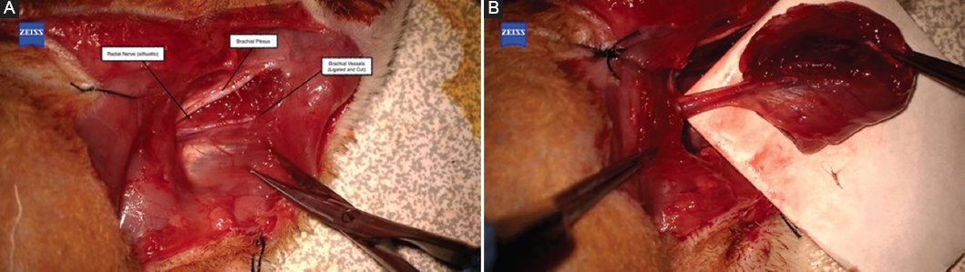
Figure 2. A: intraoperative view during the harvesting of the flap, the distal ends of the brachial vessels were ligated and the direct branches of the vessels to the muscle were preserved. B: TRICEPS brachii muscle flap prepared as an island flap based on its artery, vein and nerve.

Figure 3. The triceps brachii muscle flap was harvested as a free functional muscle flap.
Experimental groups
In our study, the left forelimb triceps brachii muscle flaps of rats in Group 1 were harvested as a pedicled flap. Then, the nerve branches innervating the muscle were cut at a distance of 3 mm from the muscle and coaptated to its original stump using 10-0 nylon suture material. The mean diameter of the nerve was found to be 0.58 mm, and the epineural neurorrhaphy technique was applied with five stitches. The distal and proximal ends of the isolated triceps muscle were sutured back to their anatomical position with 4-0 absorbable suture and kept strained.
In group 2, the triceps brachii muscle flaps of the left forelimb were harvested as a free flap, including the nerve. Then, femoral vessels and a side branch of the femoral nerve were prepared in the right groin area as the flap recipient area, and the flap inset was completed by performing end-to-end vessel anastomosis and nerve coaptation using 10-0 nylon sutures (Fig. 4). The mean diameter of vessels was measured 0.63 mm for artery and 0.66 mm for vein. Six stitches were placed for each artery anastomosis, while five stitches were applied for vein anastomosis. Then, the triceps muscle was sutured to the anterior abdominal wall and the femoral muscle with 4-0 absorbable suture for fixation and kept strained. After the surgical procedures, the incision lines were closed with 4-0 non-absorbable suture. At the end of surgery, heparin at a dose of 200 U/kg was injected subcutaneously once in the free flap group.8 All procedures were performed by the same surgeon. The animals were kept at a temperature of 21-23°C on a 12/12 h light/dark cycle (lights on at 8:00 am, lights off at 8:00 pm.). They were fed normal rodent chow in their isolated cages (600 mm × 400 mm × 200 mm). Post-operative analgesia was provided with 100 mg/kg paracetamol mixed in drinking water for 5 days. All rats were kept single in cages for 3 months.

Figure 4. An intraoperative view during free flap transfer, nerve coaptation has been completed, and artery and vein anastomoses will be done.
Outcome evaluations
At the end of 3 months of follow-up period, all animals underwent the operation under sedoanalgesia, and the triceps brachii muscle flaps and nerves that were located in the posterior arm in Group 1 and in the groin area in Group 2 were dissected from the surrounding tissue and revealed. For electromyography examination, a recording electrode was inserted into the muscle, and a ground electrode was inserted into the surrounding tissues. A standard stimulating electrode delivered current to the nerve. Pulse duration was fixed at 1 ms at a constant current from 10 μA up to 10 mA. A single shock was delivered for each trial, and muscle action potentials (MAP’s) were recorded.9 The presence or absence of reinnervation was determined with the MAP’s obtained in the muscles.
After electrophysiologic evaluation, animals were sacrificed, and biopsy samples were obtained from muscles and nerves for histopathologic analysis. The samples were fixed in 10% neutral formalin solution for 24-48 h, and then sample pieces were taken macroscopically from the muscles and microscopically from the distal part of the coaptation line for the nerves. After embedding, paraffin blocks were sectioned at 4-5 μm thickness with a microtome for Hemotoxylin and Eosin, Masson trichrome (MTC), and Luxol fast blue (LFB). MTC and LFB staining were performed with the Dako Artisan Link Pro fully automated histochemistry device. H&E and histochemical-stained slides were evaluated under a light microscope by a single pathologist.
In the microscopic examination, the inflammation density, vascularization, fibrosis, edema, fascicular organization dysfunction, axonal degeneration, myelinization and axonal density were evaluated for nerves. In addition, axons in the peripheral nerve were counted in each mm2 area. In the evaluation of muscle specimens, inflammation density, vascularization, fibrosis, muscle atrophy, necrosis and edema were evaluated.
Statistical analysis
Statistical analysis was performed using the Statistical Package for the Social Sciences 24.0 (USA). Data were evaluated for eligibility for normal distribution analysis. An analysis of variance test was used for the analysis of normal variables. Chi-squared, Fisher’s exact and Mann-Whitney U tests were used for binary comparisons and p < 0.05 was considered a significance level.
Results
At the end of 3 months period, no animal loss was observed. Wound healing was favorable in each of them, and no self-injury was seen either. Flexion contracture and limitation of extension of the left forelimb were common in the rats due to the loss of the triceps muscle. Because the muscle was restitched to its original localization, findings related to the functional disability were presented as milder in Group 1. However, it did not affect their walking and reaching for food in both groups.
According to the results of electrophysiologic evaluation, MAPs, which show the viability and contractibility of the muscles and neural healing, were recorded in the muscles with a single stimulus applied to the nerve in Groups 1 and 2. The mean MAP obtained in Group 1 was 12.475 mV, whereas this number was 12.964 mV in Group 2. There was no statistically significant comparison regarding nerve recovery.
In the histological examination, neural and muscle tissues were evaluated separately (Table 1). Vascular structures and nerve fibers (axonal density) were counted at ×200 magnification: +1, +2, and +3 indicated the presence of 0-5, 6-10, and > 10 structures, respectively. For uncountable parameters of nerve tissue (edema, disorganization, myelination, fibrosis, and inflammation) and muscle tissue (inflammation, fibrosis, muscle atrophy, and edema): +1 indicated mild, +2 indicated moderate, and +3 indicated severe. The parameters of axonal degeneration and myelinization in nerve tissue, and necrosis in muscle tissue, were evaluated as “1” for existence and “0” for absence.
Table 1. Results demonstrate the comparison of histological examinations between the groups in both the nerve and muscle tissues
| Histological evaluation | Group 1 | Group 2 | p |
|---|---|---|---|
| Neural tissue Inflammation Vascularization Fibrosis Edema Fascicular organizational dysfunction Axonal degeneration Myelinization Axonal density (×100 in a HPF) | 1.21.20.60.60.80.20.6100 | 11.61.20.61.80.60112 | 0.3170.2210.2120.1520.1000.5240.038*0.401 |
| Muscle tissue Inflammation Vascularization Fibrosis Muscle atrophy Necrosis Edema | 0.41.20.60.80.20.6 | 0.81.61.21.80.60.6 | 0.005*0.070.2140.0540.9160.476 |
|
* Indicates the statistically significant results. p < 0.05 was considered a significance level. |
|||
The signs of neural regeneration were obvious in both of the groups. The comparisons of axonal degeneration, axonal density and fascicular organizational dysfunction between the groups were not found statistically significant (p = 0.524; p = 0.401 and p = 0.100, respectively). However, myelinization was found significantly more in Group 1 by the Pearson’s Chi-squared test (p = 0.038). Moreover, the other histological evaluation parameters, such as inflammation density, vascularization, fibrosis and edema, were parallel with neural healing in both of the groups (Fig. 5).
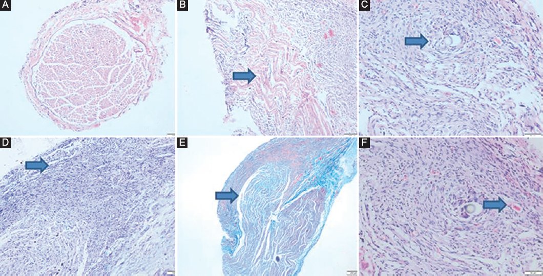
Figure 5. Microscopic examination of neural tissue. A: normal neural section and myelinization (Hematoxylin and Eosin [H&E]- × 100). B: edema (H&E- × 100). C: inflammation (H&E- × 200). D: fascicular disorganization of the peripheral nerve (Luxol fast blue- × 100). E: fibrosis (MTK- × 40). F: vascularization (H&E- × 200).
Muscle atrophy, one of the main parameters for functionality, was not found significantly different in the tests (p = 0.054) (Fig. 6). There was no flap failure, so that necrosis observed in the histopathological evaluation was at the cellular level rather than macroscopic. Inflammation was revealed more in Group 2, and the difference between groups was statistically significant (p = 0.005).

Figure 6. Microscopic examination of muscular tissue. A: focus of intense inflammation in muscle tissue (Hematoxylin and Eosin [H&E]- × 40). B: abscess area in muscle tissue (H&E- × 200). C: intense inflammation, focus of plasma cells (H&E- ×400). D: foreign body reaction (H&E- × 200). E: vascularization (H&E- × 100). F: lipomatous atrophy (H&E- × 40). G: fibrosis and atrophic changes (MTK- × 40). H: fibrosis, lipomatous changes and atrophy (MTK- × 40).
Discussion
Experimental flap models are usually defined by adapting the flaps used in the clinics to animals.10 On the other hand, the human triceps brachii muscle flap is not a commonly used flap due to the functional deficits it may cause.11 For this reason, the use of the rat triceps brachii muscle flap model defined in our study is unusual for researchers. However, it can be considered a good choice for the reconstruction of the defects in selected cases, such as covering of amputation stumps. It can also be preferred as a free fillet flap in patients with high-level upper extremity amputations.
Microsurgical training of muscle flaps is one of the crucial parts of advanced microsurgery courses. The artery, vein and nerve should be repaired meticulously to obtain viable and functional muscle after microsurgical transfer, which makes muscle flap transfer more challenging. In this study, we described a novel muscle flap in the upper extremity of the rats, which was not considered one of the muscle flap donor areas before. During the microsurgical training, increasing the number of donor areas will contribute to the use of the rats in different ways. Besides, novel injury models can be created with further studies according to the application methods of the triceps muscle flap.
Triceps brachii muscle flap harvesting causes the sacrifice of the brachial artery and vein during the procedure. Although this might suggest that the perfusion of the limb would be disturbed and a process of necrosis or autophagy would be initiated, none of the animals showed any complications related to the disruption of perfusion of the upper limb. Therefore, it can be suggested that the brachial artery can be sacrificed without disrupting limb perfusion in rats. In previously described free flaps and transplantation models, it was determined that ligating the main artery and vein of the extremity does not cause circulatory disturbance. This may be associated with the fact that collateral circulation provides effective perfusion in rats.12
Transfer of the muscles as a functional unit is not one of the frequently applied microsurgical training methods because of the complexity and requirement for supermicrosurgery. Usually, training programs of both basic and advanced microsurgical courses do not include the FFMT. The rat gracilis flap was used as an FFMT transfer model in a previous study. However, the flap was not separated from the main supplying vessel, and only the nerve was cut and repaired in that study.9 It was not possible to transport the flap to a distant body site because the flap was not separated from its vessels. In our study, the triceps flap was transported to function as both a pedicled and a free flap. With this contribution to the literature, the triceps muscle flap emerges as an FFMT model to be used in future studies. Besides, comparison between the groups was not statistically significant regarding electromyographic results, which increases the success rate of the FFMT procedure. It was also compatible with the result of mild/moderate muscle atrophy, which was seen in both of the groups.
During experimental studies, it was observed that the triceps muscle is supplied segmentally by multiple branches from the brachial vessels. If we classify this newly defined muscle flap according to the Mathes-Nahai classification, it is compatible with a Type 4 muscle flap category, which is the most difficult and unreliable type to harvest and use. This problem was overcome by harvesting the flap based on the main vessel supplying the segmental branches. On the other hand, researchers can also elevate this muscle over a single segmental vessel as a segmental muscle flap.
The fact that no necrosis was observed in any animal that underwent flap elevation and that there was no vascular variation that would affect flap harvesting during surgery can be interpreted in favor of the flap having a reliable and constant anatomy. Similarly, the absence of any complications in the donor area that prevented the continuation of the experiment is a sign that the flap is reliable as an experimental model. Besides the free flap concept, it enables functional muscle transfer, which should also be kept in mind during high-level upper extremity injuries. The reposition of the triceps muscle can be considered in patients with severe contractions in extensors and/or dysfunctional flexors, especially those who have accompanied injury in the thoracodorsal root (latissimus dorsi muscle is unavailable).
In Group 1, it was demonstrated that the triceps flap was elevated as an island flap, and this flap was supplied by the brachial artery. Moreover, in Group 2, it was shown that this flap could also be used as a free flap. According to the results obtained in the subsequent evaluations, no histopathologic and electrophysiologic difference was found between the two groups in terms of nerve healing and integrity and function of the muscle flap. Accordingly, the flap showed similar survival and functional integrity when used both as a free flap and as a pedicled flap.
Conclusions
We suggest that the rat triceps brachii muscle flap can be used as a muscle flap model. The flap can be harvested either as a pedicled flap or a free flap. Moreover, it can be used in functional muscle transfer by including the nerve if the design of the study requires. By describing a novel muscle flap in the upper extremity, we hope that it will contribute to the experimental microsurgical training, providing a new donor area. Clinical applicability of the free/pedicled triceps brachii muscle flap can be considered in selective cases.
Acknowledgments
Special thanks to Dr. IBK for her efforts on the English editing process.
Funding
The authors declare that they have not received funding.
Conflicts of interest
The authors declare no conflicts of interest.
Ethical considerations
Protection of humans and animals. The authors declare that the procedures followed complied with the ethical standards of the responsible human experimentation committee and adhered to the World Medical Association and the Declaration of Helsinki. The procedures were approved by the institutional Ethics Committee.
Confidentiality, informed consent, and ethical approval. The authors have followed their institution’s confidentiality protocols, obtained informed consent from patients, and received approval from the Ethics Committee. The SAGER guidelines were followed according to the nature of the study.
Declaration on the use of artificial intelligence. The authors declare that no generative artificial intelligence was used in the writing of this manuscript.
All procedures performed in studies involving animals were in accordance with the ethical standards of the institution at which the studies were conducted, and ethical approval was obtained from Necmettin Erbakan University, KONUDAM Experimental Medicine Research Center, with the number 2021-058.
