

Introduction
Minimally invasive surgery (MIS) is a technique that allows surgical procedures to be performed using natural pathways or small incisions in the skin to access the human body organs. This technique requires the surgeons to use a special visualization system.1 MIS procedures are increasingly important to the scientific and medical community due to their advantages in reducing complications, enabling quick patient recovery, resulting in less painful post-operative periods, and leaving fewer esthetic marks, compared to other surgical treatments.1,2 However, they require a demanding training process for surgeons.3
In many MIS procedures, observation is done on a high-resolution two-dimensional (2D) flat monitor. This is achieved by inserting an endoscope with a special arrangement of lenses and optical fiber, which transmits light generated by an external source to illuminate the cavity where the procedure is performed (abdominal, thoracic, joint, etc.).2,4 Since the projected image is in 2D, the surgeon loses the sense of depth, which complicates some operational tasks.4 Therefore, long training periods are necessary for surgeons to infer the depth of the operative field.4–6
Advances in three-dimensional (3D) visualization systems represent an opportunity to improve operative processes. This technology has been applied in surgical procedures,7 with evidence of advantages over traditional 2D visualization.4,8,9 Thus, to regain the sense of depth in MIS, comparative studies of 2D and 3D vision systems in laparoscopic training have been conducted,4,10–12 concluding that the depth vision system allows shorter learning times and greater comfort during surgery.10,13 Moreover, depth perception helps perform laparoscopic tasks more precisely,12 which could prevent post-operative complications.
Tests have been conducted with stereoscopic and autostereoscopic visualization systems,14 focusing on the technology’s usage mode. Stereoscopic displays require the user to wear or put on an element on their head, such as glasses, whereas autostereoscopic systems do not require any aids for 3D visualization.15 However, stereoscopic systems have greater commercial application, generally incorporating image capture with a dual-lens system16,17 and subsequently processing and displaying it on a monitor capable of reproducing this type of video signal.18
Significant benefits have been found in 3D visualization systems in various medical areas, such as stereopsis for MIS surgical training.19 In addition, comparative advantages have been identified in surgeon training with 3D visualization systems over 2D systems, including improved image quality, shorter average operative time, and reduced stress.20–22
Among the companies offering stereoscopic solutions for laparoscopy is Olympus, which claims that using a 3D visualization system improves the surgical experience for medical staff, increases surgeon precision, and shortens learning time.23 Other companies offer 3D monitors that enhance depth perception using active or passive glasses, such as Sony,24 Panasonic,25 Philips,26 and Eizo.27 Furthermore, manufacturers of head-mounted displays (HMDs) applicable to MIS, such as eMagin, provide an immersive 3D experience.28
In summary, and based on the literature, there is evidence that using 3D visual feedback in surgeon training at different experience levels has advantages over 2D visual feedback, along with the complement of commercial applications for professional practice in MIS. However, there is a lack of more definitive comparative studies, specifically between stereoscopic 3D visualization systems for training in laparoscopic surgery (abdominal MIS). Therefore, this study aimed to determine which 3D visualization system, among passive 3D glasses, active 3D glasses, and HMD, would achieve better results in an MIS training exercise named peg transfer. The variables used to compare the systems were: time elapsed to complete the exercise, the number of errors made during the exercise, and a subjective evaluation of image quality and ergonomics of the visualization device.
Methods
Description of the systems
To compare the 3D visualization systems, the peg transfer exercise was chosen from the fundamentals of laparoscopic surgery (FLS) training standard. This exercise allows for working in multiple spatial planes and performing ambidextrous coordination, enabling analysis of the effect of depth perception. The peg transfer task involves moving six rings from an arrangement of bars on the left side to bars on the right side, transferring them from one hand to the other while elevated, and then returning the six rings to the left side, regardless of the transfer order. As shown in figure 1, a custom Pelvitrainer box was designed to be large enough to contain a Sony 3D video camera with dual full HD lenses, a white LED lighting system, and the training exercise. Two Johnson and Johnson laparoscopic forceps were introduced by the upper access points of the Pelvitrainer for the participants to perform the exercise, following FLS recommendations. The video camera was selected to ensure that the objects to be projected would be close to the camera lenses, guaranteeing the capture of the Pelvitrainer’s workspace without needing calibration for each test subject.
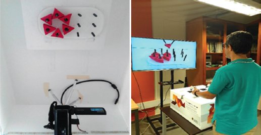
Figure 1. Upper view of the peg transfer exercise implemented in a pelvitrainer (left). Participant performing the exercise (right).
The 3D video camera was compatible with all of the following 3D stereoscopic systems that were evaluated and subsequently compared:
- − HMD. This technology is based on a multiplexed location approach, in which images are projected independently into each eye through separate channels (lenses, mirrors, and fiber optics). Vuzix900 virtual reality glasses were used in this study. These glasses have a Video Graphics Array (VGA) video input and simulate a 67” 3D screen at 9.8 feet away.
- − Passive glasses. It uses polarized multiplexing technology that divides 3D images into two images, one for each eye, by orthogonally oriented filters (linear or circular). They are known for being widely used in cinemas. For this system, a 47” LG SMART TV 3D LM67 with a high-definition multimedia interface input was used.
- − Active glasses. In this case, the “persistence of vision” effect (50 ms) is utilized to alternately and synchronously block and unblock the eyes at the same speed as the image changes, creating the sensation of continuous 3D vision. The two images for the eyes are shown at different times, at a frequency of 120 Hz or more. The device used for this system was the BenQ 3D MX666+ projector.29
During the trials, the user was ensured to perceive screens of the same size regardless of the visualization system used. To achieve this, the visualization systems were placed at different distances from the user, according to the size of the image projected by each, while maintaining the same projection ratio. As shown in figure 2, the image projected by the active system is the largest, at 53” high; the television image is the smallest, at 23” high; and the HMD image has a projection of 40.2” high, as if viewed from 118” away. Based on this last value, the location distances for the other two visualization systems were calculated. Therefore, the television was placed 67.51” from the user and the projector at 155.57.”
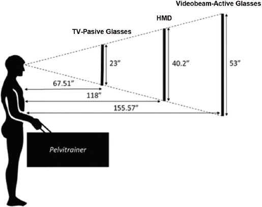
Figure 2. Distance from the participant to each of the display systems.
Protocol
The study was conducted using a randomized controlled trial to reduce cognitive biases and ensure that the different treatment groups were statistically equivalent. Since the goal was to compare three stereoscopic visualization systems (HMD, passive glasses, and active glasses) and each subject performed the peg transfer exercise with each system in a random sequence, a randomized complete block design (RCBD) was conducted.30
Following the RCBD methodology, the person performing the exercise is referred to as the subject, and each of the three visualization systems is a factor level. Randomization was applied to the order in which each test subject used each factor level, reducing the impact of confounding variables such as the person’s learning curve and fatigue level when using a particular visualization system. Controllable confounding variables in environmental conditions, such as noise, lighting, and the presence of people around the test subject, were also minimized to be consistent for each observation.
The protocol for the experiment was executed after obtaining informed consent, and each session lasted approximately 1 h. Subjects were first introduced to the exercise with a direct view of the Pelvitrainer; then, subjects performed the same training exercise but with the Pelvitrainer closed, so they did not have a direct view of the peg transfer platform. This approach helped them better understand the task and the basic handling of the laparoscopic forceps. Afterward, they performed the same exercise using 3D visualization systems: Active glasses, passive glasses, and HMDs in random order. Before and after every system (while the subject rested for 5 min), a survey of ocular systems based on the convergence insufficiency symptom survey (CISS)31,32 was performed. Visual performance data, including errors and completion times, were recorded. In addition, after completing the exercises, participants rated their experience, which included ease of use and ergonomics.
During each exercise, data on depth perception errors (missed grip, transport, or ring placement) and completion time were recorded manually and verified later using recorded videos. Parameters for comparing stereoscopic systems included:
- − The number of errors: mistakes made during the exercise were counted by reviewing video footage, following the standard method
- − Time taken to complete the FLS exercise: measured from start to finish using a stopwatch, following the FLS methodology (maximum time: 5 min)
- − Visual fatigue from using the visualization system: before and after each exercise, the CISS survey was used to assess fatigue, discomfort, double vision, eye pain, headache, drowsiness, dizziness, and blurred vision on a scale of 0-4, using a 9-question survey. A score over 13 indicates convergence issues
- − Subjective perception of image quality and ergonomics: participants rated image quality across five factors, including image blending, depth perception, image sharpness (brightness, contrast), color quality, and comfort of the glasses on a scale from 1 (Poor) to 7 (Excellent). Total scores ranged from 5 to 35.
Statistical analysis
For statistical analysis, normality tests were performed using the Shapiro-Wilk method due to its power to detect non-normality in the data and the sample size. For non-parametric analysis, the Kruskal-Wallis test for independent samples was used. Significance values were adjusted using the Bonferroni correction for multiple tests. The significant difference level considered was p < 0.05. The entire process was carried out using the Statistical Package for the Social Sciences software.
Results
Twenty-four participants without experience in the peg transfer exercise took part in the comparative test. They were aged between 18 and 35, with a mean age of 23, including 6 women and 18 men.
The results for the number of errors made by participants are shown in figure 3. A significant difference was found between the use of active glasses (median = 6, interquartile range [IQR] = 7) and the other two systems; with a p < 0.01 compared to passive glasses (median = 11, IQR = 6.5) and a p < 0.001 compared to HMD (median = 12.5, IQR = 8.5).
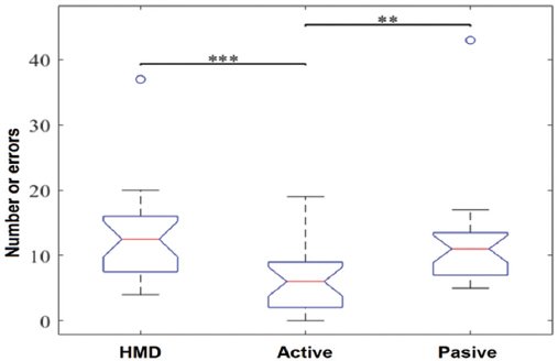
Figure 3. Comparison of the number of errors while using the visualization systems. HMD: head-mounted display. **p < 0.01 and ***p < 0.001.
On the other hand, the results for the time taken by participants to complete the peg transfer exercise are displayed in figure 4. Similar to the error variable, a significant difference was observed between the use of active glasses (median = 4.1 min, IQR = 2.1 min) and the other two systems; with a p < 0.01 compared to passive glasses (median = 6.25 min, IQR = 2.1 min) and a p < 0.001 compared to HMD (median = 7.64 min, IQR = 4.5 min).
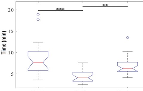
Figure 4. Comparison of the time factor according to the visualization systems when performing the exercise. HMD: head-mounted display. **p < 0.01 and ***p < 0.001.
The results of the survey on the participants’ subjective perception regarding the image quality and the ergonomics of each system (Fig. 5) show that the active glasses systems (median = 28, IQR = 8) and passive glasses systems (median = 31.5, IQR = 3) were comparable, with no significant difference (p = 0.077). However, the HMD system was perceived as lower quality (median = 20.5, IQR = 11) with a significant difference from the other two systems.
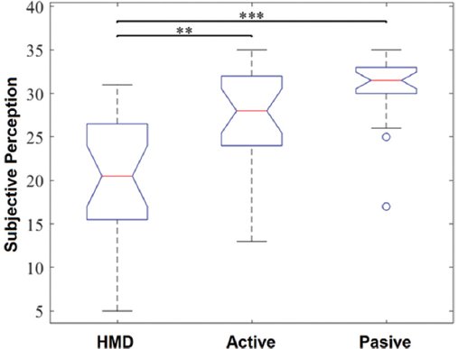
Figure 5. Comparison of users’ subjective perception regarding the visualization systems. HMD: head-mounted display. **p < 0.01 and ***p < 0.001.
Regarding the eye fatigue variable, no significant differences were observed in the results (Fig. 6). It was also determined that none of the systems caused ocular non-convergence in the users during the tests conducted with the Pelvitrainer.
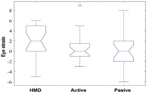
Figure 6. Comparison of the results obtained regarding eye fatigue generated by the visualization systems. HMD: head-mounted display.
Discussion
This study demonstrates that, when comparing three 3D visualization systems (HMD, passive glasses, and active glasses), the active glasses were found to be the most effective system for participants performing a basic training exercise in the FLS. This is supported by three key findings: (a) Exercises performed with active glasses were completed in less time than those with passive glasses and HMD in the peg transfer exercise of FLS, (b) exercises performed with active glasses resulted in fewer errors than those with HMD in an FLS exercise, although no significant difference was found compared to passive glasses, and (c) exercises performed with active and passive glasses resulted in a better subjective perception of image quality and system ergonomics than those with HMD in an FLS exercise.
Exercises with active glasses performed in less time
According to the results, the peg transfer exercise was completed significantly faster compared to the time taken with passive glasses and HMD, with no significant differences between the latter two in task execution time. The analysis suggests that active glasses may contribute to shorter execution times as they provide better image sharpness and depth due to alternating presentation in both eyes. In addition, they utilize the full resolution of the screen, unlike passive glasses, which use only half of it. On the other hand, the HMD used has a lower resolution due to its VGA image format, which performs less effectively compared to the digital technology of the other two systems. The Peg transfer training exercise could benefit from the use of active 3D glasses due to their increased sharpness, visual comfort, and stereoscopic precision, thus potentially improving surgical performance in manipulation tasks.
Fewer errors with active glasses
The number of errors in FLS exercises with active glasses was lower, although there was no significant difference compared to passive glasses, unlike the significantly higher errors observed with HMD. If the goal is to shorten the learning curve for trainee surgeons, active glasses are considered an appropriate technological support for such practices. Consequently, the reduction in errors with this technology would also imply less physical strain on the surgeon and improved performance in this task. While errors correlate with increased time, as found in previous studies, they are not the sole cause of procedural delays; factors such as eye fatigue, posture, motor skills, etc., must also be considered.
Achieving reduced errors and operative time during training through peg transfer exercises will be enhanced by leveraging the benefits of 3D visualization, such as improved image resolution and depth perception.
Better perception of image quality and ergonomics with active glasses
When evaluating the perception of image quality and ergonomics in wearing and using the three visualization technologies, variables such as sufficient mixing between right and left images without shadows or double vision, perception of depth, perception of object movement, color quality, and comfort of the glasses were considered critical. Any negative factor in this regard may lead to reluctance to use the system and negatively impact performance during the exercise. Results may differ, tending toward better outcomes, when performed by personnel with experience in laparoscopic surgery with 2D visualization. In addition, the effects of using high-resolution screens with 4K and 8K technologies (in both 2D and 3D, if applicable) on performance should be evaluated.
In this context, for a more comprehensive analysis regarding the performance of HMD and the other two systems, a larger number of participants is required to generalize the findings, along with longer practice times and guidance from an expert to assist in the use and resolution of learning difficulties. Furthermore, studies should be designed to estimate any retinal effects of using these new technologies to understand their impact on ocular health. Variables were assessed in users not belonging to the surgical medical field and were measured in an exercise conducted in a controlled environment rather than in a real surgical setting.
Conclusions
In the execution of the exercises, the shortest time and the lowest number of errors were obtained with active glasses, and users also benefited from perceiving depth in the image with clarity. The user’s skill, combined with the various factors evaluated, influenced the occurrence of errors in exercise execution. For the study population, the time to complete the test, the number of errors, and the system’s ergonomics were more relevant in the results than demographic variables, eye disease, or experience in using video games.
For long periods of exercise execution in this study, the weight of the vision device and the lack of peripheral vision, combined with discomfort in wearing the device, hinder the training process. Therefore, the HMD system is the least recommended for this purpose, while active glasses are the ones that yield better results.
Active glasses may allow for less fatigue in their use, better eye-hand coordination, an acceptable degree of sharpness-realism, less energy expenditure, less fatigue, and, in their analysis, the results obtained in the study reflect fewer errors, less time in task completion, and a better perception of the environment and ergonomics. Therefore, they are identified as the best option among the three devices evaluated.
As part of future tests, other FLS exercises should be covered. In addition, consideration should be given to evaluating the performance of other FLS exercises and including personnel with experience in laparoscopic surgery at different levels of expertise in the test.
Acknowledgment
The authors thank Camilo Páramo for building the Pelvitrainer.
Funding
This study was supported by the Universidad Pontificia Bolivariana, Colombia, under the project No. 088-0312-2100.
Conflicts of interest
The authors declare no conflicts of interest.
Ethical considerations
Protection of humans and animals. The authors declare that the procedures followed complied with the ethical standards of the responsible human experimentation committee and adhered to the World Medical Association and the Declaration of Helsinki. The procedures were approved by the institutional Ethics Committee.
Confidentiality, informed consent, and ethical approval. The authors have followed their institution’s confidentiality protocols, obtained informed consent from patients, and received approval from the Ethics Committee. The SAGER guidelines were followed according to the nature of the study.
Declaration on the use of artificial intelligence. The authors declare that no generative artificial intelligence was used in the writing of this manuscript.
