Introduction
Traumatic intracranial aneurysm (TIPA) is a rare high-risk subtype of cerebral aneurysm secondary to head trauma or surgery with open craniotomy.1,2 Although the incidence is low, representing only 0.15-0.40% of all intracranial aneurysms,3 it is a difficult clinical problem with high morbidity and mortality.4,5 Because of their weak walls, these lesions have a very high incidence of early rebleeding and postoperative recurrence histologically.6 Both surgical and endovascular treatments (EVTs) are challenging as these lesions involve defective vessel walls.7 Traditional therapy, including parent artery occlusion, coil embolization, and covered stent8 implantation seemed to show high rates of complications and recurrence.3
More recently, Flow–Diverter Stents have been introduced as an alternative technique for complex aneurysms, and they have shown incredible results in facilitating endovascular remodeling.9,10 However, treating percutaneous transhepatic intrahepatic portosystemic shunts (PTIPs) with flow diverters (FDs) is controversial due to the unique challenges these aneurysms present. PTIPs have fragile, thin walls lacking normal vascular layers, which increases the risk of rupture. While FDs redirect blood flow to induce thrombosis, in PTIPs, this can put additional pressure on the already fragile aneurysm, potentially causing rupture. Furthermore, the efficacy of FDs in PTIPs is not well-supported by clinical evidence, and their use requires dual antiplatelet therapy, which can increase the risk of bleeding or complicate the management of these fragile aneurysms. Other treatment options, such as surgical trapping or coiling, may be considered due to these concerns. So far, there have been only a few retrospective case series and case reports. Therefore, we conducted a meta-analysis exploring FD’s angiographic and clinical outcomes for TIPA.
Methods
Case report
An 18-year-old girl suffered head trauma 1 month ago when she fell from a bicycle. Head computed tomography (CT) showed intracranial hemorrhage with skull base fracture. However, nearly 3 days ago, swelling of the right eye, blurred vision, and pulsatile murmur developed, and he came to our hospital. Magnetic resonance imaging and digital subtraction angiography (DSA) were performed, and the diagnosis was confirmed as transcranial carotid-cavernous fistula, which drained into the superior ophthalmic vein through the cavernous sinus. The cause was considered to be the rupture of a TIPA in the cavernous segment of the internal carotid artery. Then, the FD implantation combined with coil embolization was performed under general anesthesia. Although there was still a small amount of residue on angiography immediately after the operation, the patient’s murmur disappeared. Three months after the operation, the fistula disappeared, the cavernous segment of the internal carotid artery was completely repaired, the eye symptoms disappeared, and there was no neurological dysfunction. The images were shown in figure 1.
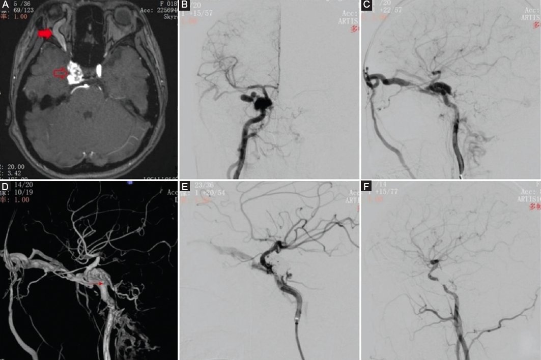
Figure 1. Case images. An 18-year-old girl with transcranial carotid-cavernous fistula (TCCF) was treated with flow diverter. A: the magnetic resonance imaging suggested a TCCF on the right side (The hollow red arrow shows the enhanced cavernous sinus, and the solid red arrow indicates the thickened ophthalmic vein). B: anteroposterior angiography of the right internal carotid artery. C: lateral angiography of the right internal carotid artery. D: 3D reconstruction image (A single arrow shows a pseudoaneurysm in the cavernous segment). E: immediate angiography after operation. F: angiography after 3 months.
Literature search strategy
A comprehensive literature search of Web of Science databases, PubMed, and Embase was conducted for studies published between 2010 and September 2022. The Preferred Reporting Items for Systematic Reviews and Meta-Analyses guidelines were followed. The keywords “aneurysm,” “intracranial aneurysm,” “pseudoaneurysm,” “PSA,” “trauma,” “traumatic,” “injury,” “flow diverter,” “flow diversion,” and “pipeline” were used in both “AND” and “OR” combinations.
Selection criteria
The systematic review used the following inclusion criteria: (1) diagnosed TIPA treated with FDs, (2) ≥ 3 patients undergoing FD implantation for TIPA, (3) clinical or angiographic outcomes were reported. Exclusion criteria were as follows: (1) Extracranial aneurysm, (2) in vitro studies and animal studies, (3) technical notes, guidelines, and review articles.
Data extraction and critical appraisal
For each study, the patient demographic data and endovascular strategies were extracted. We looked at total occlusion, procedure-related complications, progress, death, and overall satisfactory result.
Data were retrieved from the paper text, tables, and figures, and each article was examined independently by two investigators. A senior investigator reviewed the final results. Because all of the analyses were based on previously published studies, there was no need for ethical approval or patient permission.
Statistical analysis
The Newcastle–Ottawa Scale was used to assess the quality of included studies. Quality assessment was performed by two authors independently, and a third author solved potential discrepancies.
Meta-analysis was performed using the Meta package of the R software program (version 4.2.2). There are mean values for continuous variables and odds ratios and 95% confidence intervals (CIs) for dichotomous variables. Statistical heterogeneity was assessed using I2. We used a common effects model if I2 < 50%, but a random effects model if I2 > 50%, and a sensitivity analysis was conducted. It was determined that p < 0.05 was statistically significant.
Outcome measures
The term “aneurysm occlusion” was defined as complete occlusion as determined by imaging examinations (magnetic resonance angiography, CT angiography, and DSA) after the procedure. Complications were defined as any surgical treatment-related complications, including repeat bleeding, late hemorrhage, artery dissection, vessel occlusion, and brain infarction. Recurrent is defined as aneurysm enlargement or redevelopment. A good outcome was defined as 0-2 on the modified Rankin Scale or 4-5 on the Glasgow Outcome Scale.
Results
Through searches of 3 electronic databases, 369 articles were identified. After carefully examining the title or abstract, screening, and exclusion of duplicate or irrelevant articles, 7 studies with 57 patients were included. The literature search process is shown in figure 2. All studies were single-arm retrospective cohorts and had good quality. Their characteristics are summarized in table 1.

Figure 2. Flowchart of the study selection.
Table 1. Evaluation of the included studies using the modified Newcastle–Ottawa scale
| Study | Representativeness of the exposed cohort | Selection of the non-exposed cohort | Ascertainment of exposure | Demonstration that outcome of interest was not present at start of study | Comparability of cohorts based on the design or analysis | Assessment of outcome | Was follow-up long enough for outcomes to occur | Adequacy of follow-up of cohorts | total |
|---|---|---|---|---|---|---|---|---|---|
| Barburoglua et al.32 | 1 | 0 | 1 | 1 | 0 | 1 | 1 | 1 | 6 |
| Sami et al.30 | 1 | 0 | 1 | 1 | 0 | 1 | 1 | 1 | 6 |
| Chen et al.29 | 1 | 0 | 1 | 1 | 0 | 1 | 1 | 1 | 6 |
| Deng et al.17 | 1 | 0 | 1 | 1 | 0 | 1 | 1 | 1 | 6 |
| Zhao et al.18 | 1 | 0 | 1 | 1 | 0 | 1 | 1 | 1 | 6 |
| Budohoski et al.10 | 1 | 0 | 1 | 1 | 0 | 1 | 1 | 1 | 6 |
| Dogan et al.31 | 1 | 0 | 1 | 1 | 0 | 1 | 1 | 1 | 6 |
Discussion
Our meta-analysis has demonstrated that flow-diverter stents are technically feasible with a high rate of complete aneurysm occlusion and technical success for the treatment of TIPAs. The procedure has a generally favorable clinical outcome. In spite of this, the complications associated with these aneurysms were not insignificant.
TIPA is a serious type of traumatic vascular injury.11 Although the incidence is not high, its natural course prognosis is poor. The mortality rate in ruptured TICA is 30-50%.3,12 Unruptured TIPAs are more likely to rupture than non-TIPAs.13,14 Thus, definitive and immediate treatments are particularly important for the prognosis of TIPAs patients.
TPIA is fusiform and fragile, without a true neck, and is most commonly linked with dissection.12 Historically, neurosurgical treatment was mostly restricted to the sacrifice of the parent artery or bypass,15 which was heavily reliant on the existence of sufficient collateral circulation. Due to the great trauma and poor effect of open surgery, endovascular therapy has been relatively a more common treatment in recent years.3 Detachable balloons, Coil, Onyx, Stent, etc., are used in the embolization of TIPAs.11,16 These methods did not reverse the pathological process of vascular injury, and a few pseudoaneurysms recurred during the follow-up. Following, covered stents are used for EVT of TIPAs.11,17 The therapeutic strategy was also gradually changed from the initial sacrificial parent artery to the parent artery reconstruction. However, the rigid design of these systems leads to poor vascular compliance. It is often difficult to achieve full adherence to intracranial vessels, and it is relatively difficult to deliver in more tortuous vessels. In addition, its completely covered surface has the risk of blocking the perforator artery. All of these limit its application to a small number of special cases.8,18,19
FDs were introduced recently in the treatment of giant or large wide-necked aneurysms along the petrous to a superior hypophyseal segment of the internal carotid artery.20 Moreover, due to its effect on endoluminal parent vessel reconstruction, the use of FD for intracranial aneurysms has increased in popularity with new indications continuing to be reported.9,21,22 FDs have already been successfully used in a few TIPAs.23–28 Giorgianni et al.27 reported a 20-year-old man with two traumatic ACA pseudoaneurysms treated with PED that resolved after 6 months on angiography in 2015, which is perhaps one of the earliest reported in the literature. An analysis of the angiographic and clinical outcomes of TIPAs treated with FDs was conducted to determine whether FDs are feasible compared to traditional EVT. In our meta-analysis of 46 FD-treated TIPAs, the complete occlusion rate was 86.0%. Stephanie et observed one case of progression of the pseudoaneurysm in short-term follow-up, which was suggested to be associated with nonapposition of the PED on the cavernous carotid wall. Zhao11 et al. report 9 TIPAs treated with FD. They discovered 3 failures of complete occlusion at the 6-month follow-up because of no use of coils. Although coiling is a useful technique, it is risky in the setting of a recent hemorrhage and unstable TIPA capsule. All of them were completely occluded at the second follow-up, indicating that the treatment of pseudoaneurysm with FD required a longer repair time.
Although the flow-directed device has changed the local hemodynamics of the aneurysm, it does not completely isolate the aneurysmal sac and remains at risk of recurrence and rebleeding.29 Meanwhile, greater attention must be paid to operation-related complications. We assessed the complication rate as 9%. Sami30 et al., and Dogan31 et al., reported iatrogenic rupture of the distal arterioles due to a microwire during the procedure. Barburoglu and Arat32 emonstrated an individual who had an asymptomatic occlusion of the parent artery that was discovered by chance during a 7-month follow-up angiography. This patient acknowledged stopping clopidogrel for many weeks between 3- and 7-month follow-up examinations.
The present meta-analysis presents several limitations. The main limitation is the minimal number of articles analyzed due to the scarcity of literature on this subject. All of the included studies were non-comparative, single-center, retrospective case series. In addition, the baseline characteristics varied across the included studies. The lack of stratification prevented us from analyzing the effect of age, gender, FDs brand, and aneurysm location.
Conclusions
FDs are feasible and provide significant advantages in the treatment of TIPA. Nevertheless, operation-related complications are not negligible. We emphasize the therapeutic potential of FDs in TIPAs and argue that further well-designed prospective and randomized trials are needed.
Acknowledgments
We would like to acknowledge everyone for their helpful contributions to this paper.
Funding
The authors declare that they have not received funding.
Conflicts of interest
The authors declare no conflicts of interest.
Ethical considerations
Protection of humans and animals. The authors declare that the procedures followed complied with the ethical standards of the responsible human experimentation committee and adhered to the World Medical Association and the Declaration of Helsinki.
Confidentiality, informed consent, and ethical approval. The authors have followed their institution’s confidentiality protocols, obtained informed consent from patients.
Declaration on the use of artificial intelligence. The authors declare that no generative artificial intelligence was used in the writing of this manuscript.
