

Introduction
Brain trauma injury is the name given to the set of signs and symptoms produced by a mechanical alteration of the brain because of trauma.1 The ability of any penetrating object to cross the skull reaching the brain and causing primary injury is dependent on the ballistic properties, kinetic energy, mass of projectile, velocity, shape, angle of approach, the characteristics of intervening tissues, and any secondary projectiles formation such as fragments of bone or metals.2 Head gunshot injuries are increasingly encountered by neurosurgeons in civilian and urban places. Unfortunately, this is a prevalent condition in developing countries, with major armed conflicts which is not very likely to achieve a high rate of prevention.3
The number of cases related to skull gunshot injury in civilians is increasing every day.4 In countries such as the United States of America, the total costs of treating gunshot injury have been estimated at more than $16 billion/year. In addition, it generates high morbidity and mortality, leaving serious sequelae in affected patients.5
Gunshot injuries account for about 35% of head trauma deaths in people under 45 years old. They are the most fatal head injuries: about two-thirds of cases die at the scene, and, ultimately, these injuries are the direct cause of death in more than 90% of the victims.6,7 Firearm-related deaths are the second leading cause of death due to trauma in the United States; each year there are an estimated 70,000 victims of gunshot injuries, resulting in 30,000 deaths.8,9 Regarding personal injuries, in the army of the same country, 17% correspond to ballistic injuries to the cephalic pole, which results in a 0.8-day decrease in life expectancy.10 The most frequently associated age group ranges from 18 to 55 years old.11 In terms of gender, most of the victims are men. The route of entry of the projectile into the skull may vary, the most common being the right temporal 67%, followed by the mouth 16%, forehead 7%, and left temporal 7%.12
In brain injuries caused by firearm projectiles, the kinematics of the trauma are very varied. A firearm projectile injury results in permanent cylindrical cavity in the brain. The pressure waves that result in transferring kinetic energy are called “ordinary pressure waves,” which are driven radially from the projectile trajectory. These create a temporary cavity that lasts approximately 20 milliseconds as the walls of the permanent cavity first expand, then it contracts.13–15
The aim of this study is to assess whether patients who suffer brain trauma secondary to gunshot injury, who got a better Glasgow Coma Scale (GCS) score at admission, as well as a Jinnah less than 3 and a selective surgical intervention, were the most important prognostic factors for survival at 6 months. Survival was also determined in relation to age, sex, place of origin, GCS score at admission, pupillary diameter, mechanism, degree of Jinnah, treatment, and complications.
Methods
Study design
A retrospective, observational longitudinal cohort study was carried out following the Strengthening the Reporting of Observational Studies in Epidemiology guidelines.16 Permission was obtained from the hospital management to access the data from the medical records, and the research protocol was presented to the Ethics and Research Committee, where it was approved with registration number PI-2023-04.
Study population and data extraction
All records of patients over 18 years of age with a diagnosis of traumatic brain injury (TBI) due to a gunshot wound were included, from January 2019 to June 2024.
Inclusion criteria
Patients diagnosed with TBI caused by gunshot wounds and treated by the neurosurgery department. As a requirement, they must have an admission computed tomography (CT) scan with evidence of head trauma due to a gunshot wound that passed through the skull and completed a 6-month follow-up after the trauma.
Exclusion criteria
Patients with a diagnosis of death caused by gunshot wounds, gunshot wounds without cranial penetration, incomplete medical records, missing CT scans, and patients under 18 years of age. The data were collected from the medical records of patients on the following variables: sex, age, GCS score at admission, pupil diameter, mechanism, Jinnah grade, treatment, complications, and life prognosis.
Statistical analysis
The statistical analysis was conducted in three stages: descriptive analysis, subgroup comparison with statistical significance testing, and logistic regression analysis with a forest plot representation. All analyses were conducted with a significance level of p < 0.05. Descriptive analyses were used to summarize the clinical and demographic characteristics of the study population. Continuous variables (e.g., age, interaction score) were reported as mean (standard deviation [SD]), while categorical variables (e.g., sex, mechanism of injury, treatment approach) were summarized as frequencies and percentages. The overall description provided an overview of the sample before dividing it into subgroups based on survival status. Subgroup comparison: to determine whether significant differences existed between survivors and non-survivors, the sample was divided into two groups – survivors: patients with survival at 6 months and non-survivors: patients who did not survive beyond 6 months. For continuous variables, Student’s t-test was used to compare means between groups. For categorical variables, the Chi-square (χ2) test was applied. These analyses were performed using the Jamovi software to calculate p-values and identify statistically significant differences (p < 0.05). Logistic regression analysis and forest plot: to identify predictors of survival, a logistic regression model was applied. The dependent variable was survival (binary), and independent variables included those that showed significant differences in subgroup analysis (e.g., severe TBI, pupillary symmetry, treatment approach, normalized interaction score). The regression model was implemented in Python using the following libraries: pandas, NumPy, Statsmodels, and Matplotlib for creating the forest plot visualization. The forest plot illustrated the odds ratios (OR) and 95% confidence intervals (CIs) for each variable, providing a visual summary of the relative effect sizes. A significant predictor was defined as having a p < 0.05 and a 95% CI that did not include 1.
Results
Study population characteristics
Data from 65 patients diagnosed with TBI caused by gunshot wounds between January 2019 and June 2024 were collected. After excluding patients who did not meet the study criteria, a total of 44 patients were included in the analysis (Fig. 1).
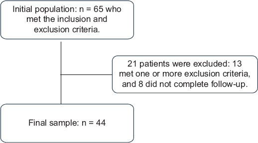
Figure 1. Flowchart of study population selection.
Of the 44 cases, 40 (91%) were men, with a mean age of 27 with an SD of 8.8 years. According to the analysis by origin, it can be determined that the majority belong to Toluca, State of Mexico, of the cases, and the rest are distributed in the State of Mexico 26 (59%). Regarding the mechanism of action, the homicidal mechanism was the most predominant, observed in 38 (86.36%) of the cases, in relation to 5 (11.36%) of the cases corresponding to the suicidal mechanism and 1 (2.27%) which represents the accidental mechanism, which is related to a wound caused by a stray bullet (Table 1).
Table 1. Clinical characteristics of the sample according to 6-month survival
| Variable | Total sample (n = 44) | Survival | p | |
|---|---|---|---|---|
| Yes (n = 25) | No (n = 19) | |||
| Age | 27.20 (8.83) | 28.60 (10.23) | 25.43 (8.5) | 0.29* |
| Sex Male Female | 40 (90.91)4 (9.09) | 23 (92)2 (8) | 17 (89.4)2 (10.6) | 0.86** |
| Mechanism Suicidal Homicidal Stray bullet/Accidental | 5 (11.36)38 (86.36)1 (2.27) | 2 (8)22 (88)1 (4) | 3 (15.7)16 (84.3)0 (0) | 0.39** |
| Initial Glasgow Coma Scale | 8.84 (4.86) | 7.8 (4.9) | 9.25 (4.49) | 0.30* |
| Brain trauma injury Mild Moderate Severe | 18 (40.91)4 (9.09)22 (50.00) | 17 (68)3 (12)5 (20) | 1 (5.2)1 (5.2)17 (89.6) | < 0.0001** |
| Glasgow outcome scale Death Severe disability Moderate Good recovery | 19 (43.18)6 (13.64)2 (4.55)17 (38.64) | 0 (0)6 (24)2 (8)17 (68) | 19 (100)0 (0)0 (0)0 (0) | NS |
| Pupils Symmetric Non-symmetric Fixed | 25 (56.82)9 (20.45)10 (22.73) | 23 (92)2 (8)0 (0) | 2 (10.5)7 (36.8)10 (52.7) | < 0.0001** |
| JINNAH I II III IV V | 20 (45.45)4 (9.09)6 (13.64)9 (20.45)5 (11.36) | 18 (72)3 (12)3 (12)0 (0)1 (4) | 2 (10.6)1 (5.2)3 (5.2)9 (47.3)4 (21) | < 0.0001** |
| Treatment Conservative Surgical Out of treatment | 13 (29.55)19 (43.18)12 (27.27) | 11 (44)14 (56)0 (0) | 2 (10.5)5 (26.3)12 (63.2) | < 0.0001** |
| Complications Hydrocephalus Cerebrospinal fluid fistula Others | 2 (4.55)2 (4.55)2 (4.55) | 1 (8)2 (16)0 (0) | 1 (5.2)0 (0)2 (10.6) | 0.135** |
| Entry site Frontal Parietal Temporal Occipital Facial | 14 (31.8)19 (43.2)4 (9.1)1 (2.3)6 (13.6) | 8 (32)13 (52)1 (4)1 (4)2 (8) | 6 (31.6)6 (31.6)3 (15.8)0 (0)4 (21.1) | 0.308** |
| Lobe Frontal Parietal Temporal Frontoparietal Temporoparietal Posterior fossa | 16 (36.3)1 (2.2)4 (9)13 (29.5)5 (11.3)5 (11.3) | 15 (60)1 (4)3 (12)3 (12)2 (8)1 (4) | 1 (5.3)0 (0)1 (5.3)10 (52.6)3 (15.8)4 (21.1) | 0.002** |
| Hospital stay 1-6 days 7-14 days 14-30 days | 20 (45.5)12 (27.2)12 (27.2) | 10 (40) 6)11 (44)4 (16) | 10 (52.6)1 (5.3)8 (42.1) | 0.011** |
| Interaction Glasgow Pupils | 4.05 (3.27) | 1.76 (1.45) | 7.05 (2.44) | < 0.001* |
| Pupillary diameter | 3.43 (2.27) | 1.24 (0.83) | 6.32 (3.04) | < 0.001* |
|
*Reported as mean (standard deviation), group comparison using Student’s t-test; **Reported as frequency (percentage), group comparison using χ2 test. |
||||
Analysis by group
The aim of the study was to compare the survival rate and identify the variables that influenced it. To achieve this, the group was divided into two subgroups. The first group consisted of patients who survived, and the second group consisted of patients who died. The overall survival rate was 25 (56.8%). The variables age, sex, mechanism of injury, site of entry, initial Glasgow Coma Score, and complications did not show statistically significant differences between the two groups (Table 1).
The neurological status, site of injury, and treatment provided were compared to assess their impact on survival, and it was found that:
NEUROLOGICAL STATUS
Severe TBI was significantly more common in the non-survival group (89.6%) compared to the survival group (20%), while mild injuries predominated in the survival group (68%) (p < 0.0001). Pupillary findings showed that symmetric pupils were observed in 92% of survivors, compared to 10.5% in non-survivors. Fixed pupils were present in 52.7% of non-survivors and absent in survivors (p < 0.0001). The Glasgow-Pupil interaction score and pupillary diameter were significantly worse in the non-survival group (7.05 ± 2.44 and 6.32 ± 3.04 mm, respectively) compared to the survival group (1.76 ± 1.45 and 1.24 ± 0.83 mm, respectively) (p < 0.001).
SITE OF INJURY
Frontal lobe involvement was more frequent in the survival group (60%) compared to the non-survival group (5.3%), while temporoparietal injuries were predominant in non-survivors (52.6%) (p = 0.002).
TREATMENT APPROACH
Surgical treatment was more frequent in survivors (56%) compared to non-survivors (26.3%). In contrast, 63.2% of non-survivors and none of the survivors were without treatment (p < 0.0001) (Table 1).
A logistic regression analysis was conducted to identify variables associated with survival in the study population. The results of the analysis were visualized using a forest plot to represent the OR and their 95% CI, observed in figure 2.
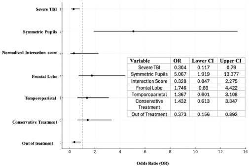
Figure 2. Forest plot of predictors of survival.
Severe TBI (OR: 0.304): severe TBI was strongly associated with lower odds of survival, indicating that patients with severe TBI were significantly less likely to survive. Symmetric pupils (OR: 5.067): the presence of symmetric pupils was a strong predictor of survival, with patients being 5 times more likely to survive compared to those with asymmetric or fixed pupils. Interaction score (OR: 0.328): lower interaction scores, reflecting greater neurological impairment, were associated with reduced survival odds. Frontal lobe injuries (OR: 1.746): injuries to the frontal lobe were associated with slightly higher odds of survival, though this finding was not statistically significant. Temporoparietal injuries (OR: 1.367): similarly, temporoparietal injuries showed a non-significant increase in survival odds. Conservative treatment (OR: 1.432): receiving conservative treatment was linked to slightly higher odds of survival, but the result was not statistically significant. Out of treatment (OR: 0.373): patients who did not receive any treatment had significantly lower odds of survival, highlighting the critical importance of timely intervention (Fig. 1).
Discussion
Penetrating head injury is the most life-threatening form of TBI, where gunshot wounds being the most common cause and the leading cause of mortality. Only 10% of patients survive to hospital, half of whom die in the emergency department and the other half often suffer significant neurological sequelae.17 The incidence of penetrating brain injuries has increased over the past two decades, mainly due to the increase in armed violence that results in 32,000-35,000 civilian deaths per year.18
Firearm-related head injury mortality is most prevalent among African American males aged 15-24 years old.19 A priori, other reviews have shown that the age groups most frequently affected by this condition tend to be between 20 and 35 years old, most of whom are men. This study shows that the most frequently affected sex is male, with an average age of 27 years old, which contrasts with international statistics, coinciding with the various studies carried out. Lower-income populations and urban communities are particularly at risk.
Making a comparison regarding the mechanism of action between the study carried out at the Jinnah Postgraduate Medical Centre, Karachi, Pakistan, it is mentioned that the homicidal mechanism was the predominant one with 51.8%, the accidental with 33.3%, and the suicidal with 14.9%; furthermore, in our hospital, the homicidal mechanism was evidenced with 86.36%, the suicidal mechanism with 11.36% and the accidental with 2.27%, both coinciding in the homicidal mechanism as the main cause of penetrating head trauma by firearm, the differences can be a result of sociopolitical arguments in a determined place and time. Furthermore, an additional study revealed 67.2% of homicide cases.20 Nearly 55% of suicides are firearm-related, with the head being the most common location.21
According to the initial GCS score in our study, we can observe that the average score at admission was 8 points, with a total of 40.91% for patients with mild TBI of which 68% survived, patients with moderate TBI were found in 9% with a 12% survival rate, and patients with severe TBI were the group with the highest proportion with 50%, of which only 20% survived. Thus, with the results obtained by the Jinnah Center in Pakistan, of which the average on admission was 9 points, in relation to the published studies in which in a multicenter study, it was observed that the average Glasgow score on admission was 8 points and 13 points in another.22,23
CT scan is the initial imaging modality of choice given its speed, wide availability, and sensitivity to detect entry and exit wounds, fractures, metallic foreign bodies, hemorrhage, and mass effect24 and its sensitivity for detecting injuries who requires surgery is close to 100%.24 In this study, non-enhanced CT scan was the base study to perform the analysis of the firearm projectile injuries, which allowed us to classify the injuries according to the Jinnah system in which we were able to obtain the following findings: Jinnah grade I lesions involving only one lobe had 45.4% of these, of which 72% of cases survived, Jinnah grade II lesions involving two or more lobes of the brain had 9%, of which 12% of cases survived, grade III lesions crossing the midline but not involving the ventricle had 13.6%, of which 12% survived, grade IV lesions crossing the midline through the ventricles of the brain had 20.45%, and grade V lesions involving the posterior fossa or brain stem had 11.36%, of which only 4% of cases survived. Therefore, we can determine that from Jinnah grade III lesions onward, the increase in mortality was substantial in the population studied. Comparing these results with the international literature, the study conducted at the Jinnah Center yielded the following results: Grade I injuries were the most common in 39% of cases, which is like the results found in our study. The highest mortality rate, 85.7%, was observed in patients with Grade V injuries, while Grade I injuries had the lowest mortality rate, 18.1%, which is like what was found in this research.25
In a study carried out in a level 1 trauma center from 2013 to 2019, which included 62 patients over 16 years of age of whom presented a bihemispheric bullet entry site, 15% survived and 71% did not survive. On the other hand, unihemispheric injury occurred in 85% of survivors and 29% of non-survivors. In relation to the affected lobes, the frontal lobe with 56% survived and 71% did not survive; the temporal lobe had 41% survived and 69% died; the parietal lobe 37% survived and 69% died; and the occipital lobe survived 19% and 20% died.26 In this study, frontal lobe involvement was more frequent in the survival group (60%) compared to the non-survival group (5.3%), while temporoparietal lesions were predominant in non-survivors. Compared with the study conducted at the level 1 trauma center, it is observed that bihemispheric condition has a worse prognosis than unihemispheric injury.
In a study carried out at the University Hospital of Monterrey, Nuevo León, an analysis of penetrating head trauma caused by a firearm projectile was carried out in which the following was observed: 80.8% of the patients were men, with an average age of 28.7 years, which presents some similarity with the results obtained in our research. Regarding the Glasgow Coma Scale on admission, it is confirmed that it is a key factor in the patient’s prognosis, related to greater morbidity and mortality when it is < 8 points, which was evidenced in the development of this research. Continuing with the analysis of the study mentioned above, 67% of the patients presented symmetrical reactive pupils upon admission, which represented a favorable prognosis; however, patients with fixed dilated pupils on admission had a 67% mortality rate. In this study, 56.82% of patients were observed with symmetrical reactive pupils, of which 92% survived at 6 months during follow-up; 20% of the patients presented with non-symmetrical pupils, of which 8% survived at 6 months of follow-up; but, again, patients with fixed pupils not reactive to light occurred in 22.73% of patients, none of whom survived.27
Management was divided into conservative and surgical. Conservative treatment consisted of positioning the patient with a headrest at 30 grades, management of cerebral edema with hypertonic solutions or mannitol, sedation with propofol in case of severe TBI, and care of wounds generated secondary to a firearm projectile, as well as strict monitoring of both neurological status and pupillary diameter every hour by trained personnel. Surgical indications include patients with asymmetric or reactive pupils, hematoma generating vector effect, or Glasgow greater than or equal to 6 points.28 Clinical factors associated with poor prognosis include Glasgow < 5 after resuscitation, dilated and nonreactive pupils, hypotension, intracranial hypertension, coagulopathy, advanced age, suicide attempt, and occipital entry wound.28 The treatment used in the management of the cases in this investigation, conservative treatment was used in 29.55% of the cases with a survival at 6 months of 44% of the cases in contrast to surgical treatment which was used in 43.18% with a survival at 6 months of 56% and the patients outside surgical management with a prevalence of 27.27% of the cases, in which no patient survived. In contrast to what is reported in the literature, the Jinnah Center yielded the following results: most cases were treated conservatively with 84.2% of cases, with a survival of 65.6% and 15.8% of patients were managed surgically, which had a survival of 72.2%, which contrasts with our results, in which there was a higher proportion of patients managed surgically. Treatment decisions may be influenced by the experience of the medical team and the policies of the center. The more aggressive surgical approach in our center may reflect a preference for early intervention in selected cases, based on the perception that surgery may be beneficial in preventing progression of neurologic deterioration as well as improving neurologic functional status.29
The Glasgow outcome scale (GOS) is a measure that classifies the patient’s condition into one of the following categories: death, severe disability, moderate disability, and good recovery intended to locate people who suffered TBI.30 In this research, patients who 38% presented good recovery, 68% of whom survived at 6 months of follow-up; in the moderate category with 4% of the patients, 8% of whom survived; regarding severe disability with 13.64% of patients with a survival of 24%; and in relation to the death category with a total of 43.18%. In contrast to Jinnah Medical Center, 30.7% of patients had a good outcome (moderate GOS and/or good recovery) and 35.9% of patients had a poor outcome (GOS death and/or severe disability). In our research, an unusual distribution of results was observed on the GOS; this may be since they presented a combination of factors that favored their recovery. These include the specific nature of the lesions, a favorable neuroplastic response, adequate surgical treatment, and a positive clinical outcome during follow-up. Individual variability in response to treatment and post-traumatic recovery means that the results of the GOS scale do not always follow a strict line according to the initial severity of the trauma but rather depend on a complex set of clinical and biological factors.
Finally, regarding the complication rate, this study showed a proportion of 86.36% of cases without complications, with a complication rate of 13.65%, the most common being hydrocephalus and cerebrospinal leak, which contrasts with the results obtained in the literature in which in Jinnah’s study, the most common complications were disseminated intravascular coagulation and hydrocephalus. Table 1 describes each of the findings obtained in this research in greater detail, which allows them to be analyzed in a more practical way.
Conclusions
This study underscores four key findings in the management of patients with penetrating cranial injuries: (1) homicidal mechanism as the primary cause of these injuries, particularly in young males, highlighting armed violence as a determinant factor in public health; (2) the importance of the GCS score and pupillary diameter upon admission as essential prognostic factors, allowing for rapid classification of injury severity and early guidance in clinical decision-making; (3) the critical role of the Jinnah classification, which helps predict survival and determine appropriate treatment, distinguishing those patients who may benefit from a conservative approach from those requiring immediate surgical intervention; and (4) the location of the injury, especially concerning the affected brain lobes, also plays a decisive role. Bihemispheric lesions have an especially reserved prognosis due to the involvement of both cerebral hemispheres, compromising vital brain functions.
These findings have significant implications for clinical practice, as they highlight the utility of prognostic factors such as the GCS and pupillary diameter in decision-making, allowing for early identification of patients who may benefit from either conservative or surgical treatment. The Jinnah classification provides a stratification tool that optimizes the management and prioritization process in trauma units.
A relevant finding from this study is that, while complications were less frequent in our cohort compared to other studies, conservative treatment showed a slightly higher survival rate than surgical treatment, although this result should be interpreted with caution due to sample size limitations. This finding paves the way for future research to validate these observations and more definitively determine the most effective strategies for treating such injuries.
Finally, the findings of this study not only have clinical implications but also reflect a profound social issue related to armed violence, underscoring the need for multidisciplinary approaches that include both improvements in medical management protocols and social interventions addressing the underlying causes of homicidal injuries. This study can serve as a foundation for improving management protocols and patient prioritization, optimizing the care of those with penetrating cranial injuries, and potentially improving clinical outcomes in this vulnerable population.
Funding
The authors declare that they have not received funding.
Conflicts of interest
The authors declare no conflicts of interest.
Ethical considerations
Protection of humans and animals. The authors declare that no experiments involving humans or animals were conducted for this research.
Confidentiality, informed consent, and ethical approval. The study does not involve patient personal data. The SAGER guidelines were followed according to the nature of the study.
Declaration on the use of artificial intelligence. The authors declare that no generative artificial intelligence was used in the writing of this manuscript.
