Introduction
Among malignancies within the gastrointestinal system, pancreatic cancer exhibits notably high mortality rates. While surgery remains the most effective treatment approach, the 5-year survival rate stands at 10-25%.1,2 Determination of resectability primarily relies on pre-operative imaging techniques such as computed tomography (CT), magnetic resonance imaging, and endoscopic ultrasound, alongside biological factors. Numerous criteria, encompassing anatomical, biological, and conditional aspects, have been proposed to assess the resectability of pancreatic tumors.3 Anatomical parameters, including invasion of the superior mesenteric artery, hepatic artery, and aberrant hepatic artery, as well as unreconstructable involvement of the superior mesenteric vein and portal vein structures, were assessed as indications for unresectable conditions. Biological factors encompass lymphadenopathies that lack pathological confirmation on imaging or elevated levels of CA 19-9 exceeding 500 U/mL. Conditional variables were evaluated in conjunction with the patient’s Eastern Cooperative Oncology Group (ECOG) score.4 Patients with elevated CA 19-9 levels, a sizable tumor on imaging, an indicative enlarged lymph node, substantial weight loss, and intense pain are categorized as high-risk individuals. Within this patient cohort, a more targeted diagnostic laparoscopy is recommended to assess resectability and ascertain the presence of micrometastasis.5
In pre-operative assessments, pancreatic ductal adenocarcinomas (PDACs) initially deemed operable based on contrast-enhanced CT scans were subsequently found to be unresectable in approximately half of the cases.6 While CT scans exhibit a high predictive value for identifying unresectable disease, their sensitivity in detecting resectable PDAC is limited. Consequently, alongside radiological imaging, biological factors are employed to evaluate the resectability of PDAC in patients.
The objective of our study is to identify novel radiological and biological variables, alongside the established criteria outlined in current guidelines, for determining the pre-operative resectability status of patients diagnosed with radiologically resectable PDAC.
Methods
Study design and patients
This research was conducted in accordance with the World Medical Association Declaration of Helsinki. An opt-out informed consent protocol was used for this study. This consent procedure was reviewed and approved by the local ethical review board, approval number (2023/4702).
This retrospective study was conducted at a tertiary care center and included data from all patients who underwent the Whipple procedure at our facility between 2010 and 2022.
Inclusion criteria
- – Tumors localized to the right side of the pancreas
- – Anatomical structures appear amenable to surgical resection based on the assessment using pre-operative CT imaging
- – Performance status ECOG 0 and ECOG 1
- – Patients with final diagnosis of PDAC.
Exclusion criteria
- – Neoadjuvant therapy
- – Patients with distant metastasis detected on pre-operative CT.
During the course of the investigation, the resectability of the tumor was assessed by two experienced surgeons. Patients were categorized into resectable or unresectable groups based on the surgeons’ judgment. Collected data included patient age; pre-operative laboratory measurements such as alanine aminotransferase, aspartate aminotransferase, γ-glutamyl transferase, total bilirubin, albumin, carbohydrate antigen (CA19-9), and carcinoembryonic antigen (CEA); lymphocyte count (103/uL); neutrophil count (103/uL); platelet count (103/uL); neutrophil-to-lymphocyte ratio (NLR); platelet-to-lymphocyte ratio (PLR); and tumor size observed on CT. The necessary data were obtained from the patients’ database. Pre-operative imaging was reviewed by a specialized radiologist to assess anatomical resectability and identify the presence of distant metastases.
Statistical analysis
The sample size for this study was determined using power analysis with G-Power 3.1 software. A power (1-β) of 0.80 and a confidence level of 95% were considered, resulting in a calculated sample size of 29 participants per group. Therefore, a minimum total sample size of 58 participants was determined for both groups. The normality of the distribution was assessed using the Shapiro-Wilk test. Non-parametric tests, specifically the Mann-Whitney U test, were employed as appropriate. Descriptive statistics, including median and interquartile range (IQR), were reported for the variables. The optimal cutoff point for tumor size, lymphocyte count, NLR, and PLR was determined using receiver operating characteristic (ROC) curve analysis. Categorical variables were analyzed using X2 analysis, and the results were presented as frequencies and percentages. Subsequently, a prospective selective multivariate logistic regression analysis was conducted to examine the variables that demonstrated statistical significance. The goodness of fit of the logistic regression model was assessed using the Hosmer-Lemeshow test. Statistical significance was defined as p < 0.05.
Results
Demographic and clinical characteristics
A retrospective analysis was conducted on the data of 159 patients who underwent laparotomy for right-side pancreatic mass in a study. Pathology results from 39 patients (24.53%) were excluded as they did not meet the criteria for PDAC. Among the remaining 120 patients included in the analysis, 30 (25.0%) were found to be unresectable during intraoperative exploration (Fig. 1). A detailed analysis of patient data is presented in tables 1 and 2. The median age (IQR) of the study population was 66 (59-73), with 84 (70.0%) being male. Moreover, the performance status of the patients who underwent surgery was categorized as ECOG0 in 39 individuals (67.5%) and ECOG 1 in 81 individuals (32.5%).
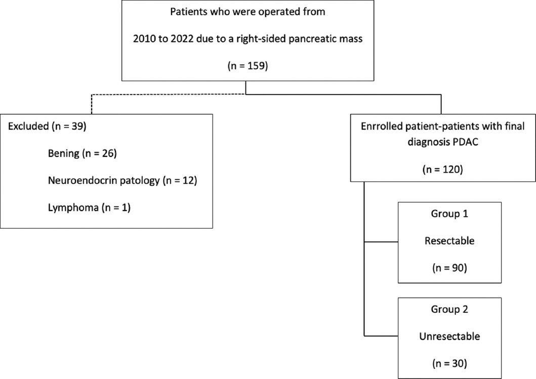
Figure 1. Patient flowchart
Table 1. Demographic and clinical characteristics of included patients
| Variables | Median (IQR) | Count (%) |
|---|---|---|
| Age | 66 (59-73) | |
| Gender Female Male | 36 (30.0)84 (70.0) | |
| Performance status ECOG 0 ECOG 1 | 81 (67.5)39 (32.5) | |
| Resectability Unresectable Resectable | 30 (25.0)90 (75.0) | |
| Morbidity (CD) I-II III-IV | 8 (6.7)97 (80.8)23 (19.2) | |
|
IQR: interquartile range; ECOG: Eastern Cooperative Oncology Group; CD: Clavien-Dindo classification. |
||
Table 2. Univariate analysis of factors predicting unresectability
| Variables | Resectability | p | |||
|---|---|---|---|---|---|
| Unresectable | Resectable | ||||
| Median (IQR) | Count (%) | Median (IQR) | Count (%) | ||
| Age, year | 74 (69-78) | 63 (58-70) | <0.001 | ||
| Gender Female Male | 9 (30.0)21 (70.0) | 27 (30.0)63 (70.0) | 0.597 | ||
| Performance status ECOG 0 ECOG 1 | 18 (60.0)12 (40.0) | 63 (70.0)27 (30.0) | 0.311 | ||
| Tumor size, cm | 4.65 (4-5.5) | 3.5 (2.9-4.5) | <0.001 | ||
| CA 19-9, U/mL | 375.95 (20-1016) | 47 (1.6-282.1) | 0.002 | ||
| CEA, ng/mL | 5.48 (3.10-8.62) | 2.5 (1-4.2) | <0.001 | ||
| Albumin, g/dL | 3.2 (2.7-3.9) | 3.3 (2.8-3.7) | 0.932 | ||
| AST, U/L | 58 (23-135) | 42.5 (21-87) | 0.231 | ||
| ALT, U/L | 57.5 (25-212) | 63 (28-122) | 0.942 | ||
| GGT, IU/L | 117 (66-453) | 178 (58-369) | 0.894 | ||
| Total Bilirubin, mg/dL | 1.15 (0.61-9.32) | 1.9 (0.9-6.4) | 0.456 | ||
| Lymphocyte, 103/uL | 1.34 (0.94-1.71) | 1.8 (1.5-2.3) | <0.001 | ||
| Neutrophil, 103/uL | 4.98 (3.76-5.45) | 4.85 (3.6-6.5) | 0.524 | ||
| Platelet, 103/uL | 250 (208-311) | 290.5 (221-336) | 0.083 | ||
| NLR | 3.46 (2.76-5.33) | 2.41 (1.81-3.87) | 0.013 | ||
| PLR | 205.17 (135-252.31) | 148.05 (108-205.83) | 0.034 | ||
| Morbidity (CD) I-II III-IV | 1 (3.3)25 (83.3)5 (16.7) | 7 (7.8)72 (80)18 (20) | 0.3980.688 | ||
|
p < 0.05 was considered statistically significant. CA 19-9: carbohydrate antigen 19-9; CEA: carcinoembryonic antigen; NLR: neutrophil-to-lymphocyte ratio; PLR: platelet-to-lymphocyte ratio; CD: Clavien-Dindo classification; ALT: alanine aminotransferase; AST: aspartate aminotransferase; GGT: γ-glutamyl transferase. |
|||||
Predictive factors for unresectable and resectable on exploration
The group of patients with unresectable tumors displayed several distinct characteristics compared to the group with resectable tumors. First, the unresectable group consisted predominantly of elderly individuals, with a median age of 74 years, whereas the resectable group had a median age of 63 years (p < 0.001). Furthermore, the unresectable group exhibited significantly larger tumor sizes, with a median value of 4.65 cm, as opposed to 3.5 cm in the resectable group (p < 0.001).
In addition, patients belonging to the unresectable group demonstrated higher levels of specific biomarkers in comparison to the resectable group. Specifically, the unresectable group displayed elevated levels of CA 19-9 (375.95 vs. 47 U/mL, p = 0.002), CEA (5.48 vs. 2.5 ng/mL, p < 0.001), NLR (3.46 vs. 2.41, p = 0.013), and PLR (205.17 vs. 148.05, p = 0.034) in comparison to the resectable group. However, it is important to note that the lymphocyte count was significantly lower in the unresectable group (1.34 vs. 1.8 103/uL, p < 0.001) (Table 2).
The ROC curve analysis was performed to determine the optimal cutoff points for tumor size (≥ 4 cm), CA 19-9 (≥ 75 U/mL), CEA (≥ 6 ng/mL), lymphocyte count (≤ 1.55 103/uL), NLR (≥ 3.2), and PLR (≥ 171). The analysis aimed to identify the threshold values at which these parameters could effectively discriminate between different groups. In addition, the specific value of CA 19-9 > 500 U/mL was also assessed and tested (Table 3).
Table 3. Validity of independent factors predicting unresectability
| Variables | Cutoff | Sensitivity (%) | Specificity (%) | AUC | 95% CI | p |
|---|---|---|---|---|---|---|
| Tumor size, cm | ≥ 4 | 76.67 | 60.00 | 0.683 | 0.576-0.791 | 0.003 |
| CA 19-9, U/mL | > 500 | 50.00 | 81.11 | 0.656 | 0.536-0.775 | 0.011 |
| CA 19-9, U/mL | ≥ 75 | 56.67 | 56.67 | 0.567 | 0.448-0.685 | 0.275 |
| CEA, ng/mL | ≥ 6 | 50.00 | 83.33 | 0.667 | 0.547-0.786 | 0.006 |
| Lymphocyte, 103/uL | ≤ 1.55 | 70.00 | 66.67 | 0.683 | 0.573-0.794 | 0.003 |
| NLR | ≥ 3.2 | 63.33 | 64.44 | 0.652 | 0.524-0.754 | 0.023 |
| PLR | ≥ 171 | 63.33 | 63.33 | 0.630 | 0.518-0.749 | 0.029 |
|
p < 0.05 was considered statistically significant. CA 19-9: carbohydrate antigen 19-9; CEA: carcinoembryonic antigen; NLR: neutrophil-to-lymphocyte ratio; PLR: platelet-to-lymphocyte ratio; AUC: area under the curve; CI: confidence interval. |
||||||
In univariate regression analyses, several factors were found to be significantly associated with the resectability status of PDAC during surgery. These factors included older age (p < 0.001), larger tumor size (p = 0.001), elevated levels of CA 19-9 (> 500 U/mL; p < 0.001, ≥ 75 ng/mL; p = 0.207), elevated levels of CEA (p < 0.001), increased NLR (p = 0.009), higher PLR (PLR; p = 0.012), and lower lymphocyte count (< 0.001) (Table 4).
Table 4. Univariate and multivariate logistic regression analysis results
| Variables | Univariate logistic regression | Multivariate logistic regression | ||||
|---|---|---|---|---|---|---|
| OR | 95.0% CI | p | OR | 95.0% CI | p | |
| Tumor size ≥ 4 cm | 4.929 | 1.915-12.684 | 0.001* | 4.152 | 1.358-12.697 | 0.013 |
| CA 19-9 > 500 U/mL | 4.294 | 1.765-10.449 | < 0.001* | 9.929 | 1.602-61.552 | 0.014 |
| CA 19-9 ≥ 75 U/mL | 1.710 | 0.743-3.937 | 0.207 | 5.238 | 0.898-30.573 | 0.066 |
| CEA ≥ 6 ng/mL | 5.000 | 2.022-12.363 | < 0.001* | 2.130 | 0.713-6.367 | 0.176 |
| Lymphocyte ≤ 1.55 103/uL | 4.667 | 1.906-11.426 | < 0.001* | 4.253 | 1.142-15.838 | 0.031 |
| NLR ≥ 3.2 | 3.131 | 1.326-7.390 | 0.009* | 1.706 | 0.439-6.634 | 0.441 |
| PLR ≥ 171 | 2.983 | 1.266-7.032 | 0.012* | 1.485 | 0.357-6.180 | 0.587 |
|
p < 0.05 was considered statistically significant. CA 19-9: carbohydrate antigen 19-9; CEA: carcinoembryonic antigen; NLR: neutrophil-to-lymphocyte ratio; PLR: platelet-to-lymphocyte ratio; OR: odds ratio; CI: confidence interval. |
||||||
In the multivariate analysis, after adjusting for other variables, the following factors remained significantly associated with the unresectable group. These factors were tumor size ≥ 4 cm (odds ratio [OR] = 4.152; 95% confidence interval [CI] = 1.358-12.697; p = 0.013), CA 19-9 > 500 U/mL (OR = 9.929; 95% CI = 1.602-61.552; p = 0.014), and lymphocyte count ≤ 1.55 103/uL (OR = 4.253; 95% CI = 1.142-15.838; p = 0.031) (Table 4).
Discussion
Currently, the evaluation of resectability in PDAC patients is based on the pre-operative criteria set by the International Association of Pancreatology (IAP).3 Treatment decisions for patients are made accordingly. However, there is a subset of patients with radiologically potentially resectable PDAC who do not receive curative treatment. One of the contributing factors is the limitations of imaging methods. Consequently, unnecessary laparotomy procedures are performed in cases where pancreatic resection is not feasible, leading to significant morbidities. Moreover, this delays the initiation of chemotherapy regimens, resulting in reduced overall survival for patients.
Recent studies conducted on radiologically potentially resectable PDAC patients have revealed that several factors, including CA 19-9, CEA, CA 125, DUPAN-2, tumor size, weight loss, and tumor location (specifically left-sided tumors), can serve as predictive indicators of unresectability.7–10
In our study, we observed that tumor size, CA 19-9 levels, CEA levels, lymphocyte counts, NLR, and PLR exhibited statistically significant associations in univariate regression analyses. However, after adjusting for potential confounders, we found that tumor size ≥ 4 cm, CA 19-9 levels exceeding 500 U/mL, and lymphocyte counts ≤ 1.55 × 10³/uL emerged as independent risk factors for resectability.
CA 19-9 is extensively investigated as a tumor marker for the diagnosis and prognosis of pancreatic cancers. In our study, we determined a cutoff value of 75. In addition, we examined the recommended value of 500 U/mL by the IAP. An elevated CA 19-9 level of 500 U/mL was found to increase the risk of the tumor being unresectable approximately tenfold. Van Manen et al.7 suggested a cutoff of 305 U/mL for CA19-9, which resulted in an OR of 2.58 (95% CI 1.30-5.14; p = 0.007). Zhang et al. reported that a CA 19-9 level exceeding 353.3 U/mL predicted non-resectability in pancreatic cancer patients who underwent laparotomy.11 Conversely, the study by Benke et al. emphasized that CA 19-9 should not be solely relied upon as a safe marker for determining resectability in surgical decision-making.12
Our study’s observed low cutoff value is likely attributed to the inclusion of exclusively right-sided pancreatic tumors. This selection may have influenced the results, as left-sided pancreatic tumors exhibit a higher prevalence of distant metastasis and locally advanced stage upon diagnosis.13
It is widely recognized that pancreatic head adenocarcinomas often coincide with elevated bilirubin levels. Consequently, CA 19-9 measurements may not be suitable for assessing patients with jaundice. A correlated study demonstrated that a CA 19-9 value exceeding 178 U/mL can serve as a reliable indicator of unresectable disease in patients experiencing jaundice.14
The enlargement of pancreatic tumors, similar to other malignancies, has a notable impact on prognosis, overall survival, and the likelihood of resectability. A study conducted by Chiang et al. indicated that pancreatic adenocarcinomas with a tumor size exceeding 4.8 cm are more prone to being unresectable. The authors also emphasized the importance of evaluating clinical characteristics of the tumor in conjunction with radiological imaging.15 Another study reported a higher probability of unresectability in pancreatic adenocarcinomas with a tumor size of 3 cm or greater.8 In our study, we found that cases with a tumor size of 4 cm or larger had an approximately fourfold increase in unresectability. It is important to note that the distinguishing factor of our study was the standardized patient group consisting exclusively of right-sided pancreatic tumors.
Immunosuppression plays a significant role in the advancement of malignancies, including pancreatic cancer. Both lymphopenia and a high NLR serve as indicators of an unfavorable prognosis, indicating the presence of advanced-stage disease.16 A study further highlighted that an NLR exceeding 3.74 independently increases the risk in cases of unresectable PDAC.17 In our study, although NLR did not emerge as an independent risk factor, lymphopenia demonstrated predictive value for identifying unresectable PDAC.
Limitations of the study
A limitation of the study lies in the relatively small cohort of patients with unresectable conditions. By broadening the study population, we can effectively elucidate the risk factors associated with resectability in a more definitive manner. Furthermore, this expansion will facilitate the development of nomograms, enabling accurate prognostic predictions and individualized treatment strategies.
Conclusions
In cases of radiologically assessable PDACs, the incorporation of variables such as CA19-9 > 500 U/mL, lymphocyte ≤ 1.55 103/uL, and tumor size ≥ 4 cm can enhance the precision of resectability assessment. This refined evaluation aids in minimizing unwarranted laparotomies. Moreover, it is prudent to contemplate exploratory intervention following neoadjuvant therapy when there is concurrent manifestation of all three specified prognostic indicators. This counsel is underpinned by the plausible prospect of encountering non-resectable pathologies during the course of diagnostic intra-abdominal scrutiny.
Funding
The authors declare that they have not received funding.
Conflicts of interest
The authors declare no conflicts of interest.
Ethical considerations
Protection of human subjects and animals. The authors declare that no experiments on humans or animals were performed for this research.
Confidentiality, informed consent, and ethical approval. The authors have obtained approval from the Ethics Committee for the analysis of routinely collected and anonymized clinical data; therefore, individual informed consent was not required. Relevant ethical recommendations have been followed.
Declaration on the use of artificial intelligence. The authors declare that no generative artificial intelligence was used in the writing or creation of the content of this manuscript.
