

Introduction
Spinal fusion is among the most frequently performed surgical procedures worldwide, particularly by spine surgeons. The Agency for Healthcare Research and Quality estimates that over 488,000 spinal fusion surgeries are performed annually in the United States, accounting for 3.1% of all surgical procedures.1 Spinal fusion is indicated for various conditions, including degenerative disc disease, disc herniation, vertebral fractures, and spinal deformities. This procedure involves fusing two or more vertebral segments using screw-rod systems, plates, cages, or a combination of these techniques.2
To optimize surgical outcomes, extensive research has focused on surgical techniques, fusion materials, and adjunctive therapies such as grafting, electromagnetic stimulation, and pharmacological agents. Insufficient bone fusion can result in complications such as pseudoarthrosis, non-union, instrumentation failure, and iatrogenic deformities, significantly diminishing patients’ quality of life.3 Bone grafting remains a cornerstone of fusion surgery, with options including autografts, allografts, xenografts, and synthetic materials.4 In addition, biological agents such as RANKL inhibitors (e.g., denosumab), parathyroid hormone analogs, growth factors (e.g., Insulin-like growth factor, fibroblast growth factor, and B-epidermal growth factor), and sonic hedgehog genes have demonstrated efficacy in promoting bone healing.5,6
Equally critical is understanding factors that hinder bone fusion. Metabolic disorders (e.g., diabetes mellitus and osteoporosis), vitamin deficiencies, vascular issues (e.g., anemia and hypoxia), mechanical factors (e.g., inadequate immobilization and instrumentation failure), and lifestyle factors (e.g., tobacco and alcohol use) have been shown to impair bone healing.7 Furthermore, psychological factors such as depression and distress are known to affect wound healing adversely.8 However, the impact of depression on spinal fusion remains unexplored.
Katz’s unpredictable chronic mild stress (UCMS) model, developed in the 1980s, serves as a widely used model for studying depression.9 Given that bone fusion is a form of wound healing, this study aims to investigate the effect of psychological distress and depression on spinal fusion using a rabbit posterolateral fusion model.
Methods
Setting and ethics committee
This study was conducted in the Department of Experimental Studies at a university hospital with ethics approval was obtained from the same institution (approval number: 776). The experiment was designed to last for a total duration of 8 weeks.
Selection criteria for the study and grouping of the animals
A total of 12 male New Zealand rabbits, aged 8 months with a mean weight of 3250 g, were selected for the study. Each rabbit was housed individually in separate cages. The animals were randomly divided into two equal groups: an experimental group and a control group, each consisting of six rabbits.
The control group was maintained under standard laboratory conditions, with ad libitum feeding, a 12-h light-dark cycle, a controlled room temperature of 20-24°C, and 60% relative humidity. In contrast, the experimental group was subjected to the conditions of UCMS, which will be described in detail later. All rabbits underwent identical surgical procedures as part of the study protocol.
Exclusion criteria
To ensure adherence to ethical standards and animal welfare while minimizing potential bias in the results, the following exclusion criteria were established:
- − Rabbits that died before the completion of the 8-week study period, either preoperatively, intraoperatively, or postoperatively
- − Rabbits that failed to eat or drink adequately during the study
- − Rabbits that were unresponsive to external stimuli
- − Rabbits deemed unsuitable for the study by the attending veterinarian.
Fortunately, no adverse events occurred during the study, and all rabbits met the inclusion criteria, enabling the collection of optimal data.
Anesthesia
In an operating room environment with air exchange every 5 min, a controlled temperature of 22° C, and 60% humidity, pre-anesthesia was administered intramuscularly using 8 mg/kg xylazine (Rompun 2%©, Bayer AG, Germany). Once the animals were sedated, anesthesia was induced with 40 mg/kg ketamine (Ketasol 10%©, Richter Pharma AG, Austria) administered intraperitoneally. Proper anesthetic depth was confirmed by the veterinary team through the absence of a pain response to foot pinching. To prevent corneal dryness during the procedure, eye drops (Viscotears©, Alcon Pharma, Switzerland) were applied.
First surgery
Once anesthesia was confirmed, the rabbits were positioned prone. The surgical field was shaved and thoroughly cleansed with an antiseptic solution. To minimize intraoperative bleeding and post-operative pain, 2 cc of prilocaine hydrochloride (Pricain 2%©, Polifarma İlaç, Turkey) was administered subcutaneously and intramuscularly into the surgical area. The left and right iliac crests were palpated, and a 2 cm skin incision was made parallel to the left iliac crest. The area was exposed through blunt dissection, and 2 g of bone was harvested from the left iliac crest using a rongeur (Fig. 1A).

Figure 1. A: harvesting of the iliac bone graft. B: anatomic orientation and visualization of the L5-L6 intervertebral space. C: inserting the bone grafts and closing the wound.
A midline skin incision was then made from L4 to S2 to access the rabbit spine. The paravertebral muscles were dissected, exposing the posterior spinal elements (Fig. 1B). As no instrumentation was planned, the left L5 and L6 facets and transverse processes were decorticated using a rongeur to create a stable environment for fusion. The harvested iliac crest bone graft was placed in the prepared area, and the wound was closed with 3/0 silk sutures (Fig. 1C).
Postoperatively, the rabbits were monitored until full recovery from anesthesia. All animals retained normal motor function following the procedure. Wounds were inspected and cleaned daily. For pain management, the rabbits received 2 mg/kg carprofen (Rimadyl XL©, Zoetis, USA) for 5 days.
Applying the UCMS model
The rabbits in the control group were monitored under standard laboratory conditions for 8 weeks. Those in the experimental group were subjected to the UCMS model, implemented as follows:
Day 1
A tub was filled with tap water at 24° C to a depth of 1 cm. The rabbits in the experimental group were placed in the tub for 4 h (Fig. 2). Afterward, they were removed, dried, warmed under a 37° C heating lamp, and returned to their cages.

Figure 2. A water tub in which the rabbits were kept for 4 h.
Day 2
The standard 12-h light and dark cycle was altered. The animals were kept in complete darkness for 24 h.
Day 3
The light-dark cycle was disrupted again, with the lights left on continuously for 24 h.
Day 4
A 5 cm box was placed under one side of the shelf holding the cages, tilting the cages to create an inclined position. The rabbits remain in this setting for 4 h (Fig. 3).

Figure 3. Elevation construct with a 5 cm box providing an inclination of the cages.
Day 5
The rabbits were confined to a cage typically designed for rats, restricting their movement and inducing a claustrophobic environment for 4 h (Fig. 4).

Figure 4. Putting the rabbits in a rabbit cage to achieve claustrophobia.
Day 6
Each rabbit was placed in another rabbit’s cage for 4 h before being returned to its own.
Day 7
No stress-inducing intervention was performed.
To prevent habituation, the cycle was altered each week. During the experimental period, the rabbits exhibited behavioral changes, including reduced food and water intake and decreased activity, which are consistent with the core clinical symptom of depression: anhedonia.
Second surgery
After 8 weeks, all animals from the experimental and control groups were sedated with 8 mg/kg intramuscular xylazine (Rompun 2%©, Bayer AG, Germany) and euthanized via intracardiac injection of 100 mg/kg potassium chloride. The midline incision was reopened, and macroscopic observations in some rabbits revealed fusion between the harvested bone graft and the facet and transverse processes.
The vertebral segments were carefully dissected free of surrounding soft tissue and removed en bloc using a wire saw. The extracted vertebrae were palpated and assessed for evidence of fusion by manually applying flexion and extension maneuvers. The results were categorized as “fusion” or “no fusion” (Fig. 5).
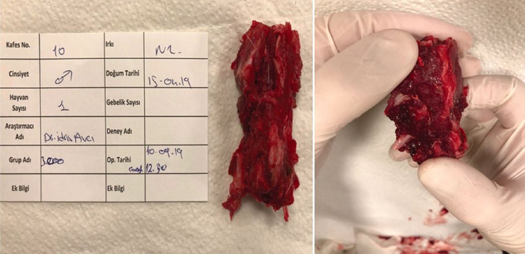
Figure 5. Manual palpation and applying flexion-extension movements on the extracted spine segment.
Subsequently, the vertebrae were preserved in formaldehyde and transported to the radiology department. Anterior-posterior images were obtained using a fluoroscopic C-arm (Ziehm Solo©, Ziehm Imaging, Germany). Radiographic evaluation focused on signs of fusion. A scoring system was applied: vertebrae without callus formation or sclerosis, indicating no evidence of fusion, received 0 points. Vertebrae displaying callus formation, an early marker of fusion, were assigned 1 point. Complete fusion, characterized by sclerosis, received 2 points (Fig. 6).

Figure 6. Radiological X-ray images of the extracted spine segment. A: no sign of fusion = 0 points. B: callus formation = 1 point. C: sclerosis formation = 2 points.
Results
Statistical analysis
The results were analyzed using the Z-test to compare categorical measurements between the two independent groups. Numeric and percentage values were applied as complementary statistical tools for categorical variables. Statistical analysis was performed using the Statistical Package for the Social Sciences v.24 (IBM, USA), and p < 0.05 was considered statistically significant.
Manual palpation results
Bone fusion was evaluated using two methods. The first involved manual flexion-extension maneuvers performed on the en bloc extracted vertebral segments. In the experimental group, subjected to the UCMS model, fusion was observed in only 2 out of 6 rabbits (33.3%). In contrast, in the control group, which was not exposed to stress, 5 out of 6 rabbits (83.3%) exhibited fusion.
The results from the manual palpation method indicated that UCMS had a significant negative effect on bone fusion compared to the control group (p = 0.045) (Table 1a–c).
Table 1a. Evaluation of fusion after manual palpation. Evaluation of fusion in the experimental group
| Rabbit number | Fusion |
|---|---|
| 1 | No |
| 2 | No |
| 3 | Yes |
| 4 | No |
| 5 | No |
| 6 | Yes |
Table 1b. Evaluation of fusion in the control group
| Rabbit number | Fusion |
|---|---|
| 1 | Yes |
| 2 | Yes |
| 3 | No |
| 4 | Yes |
| 5 | Yes |
| 6 | Yes |
Table 1c. Comparison of fusion between the experimental and control groups
| Experimental group | Control group | Total | z | p | |
|---|---|---|---|---|---|
| Fusion | 2/6 = 0.33 | 5/6 = 0.83 | 7/12 = 0.58 | 2.020 | 0.045 |
| No fusion | 4/6 = 0.67 | 1/6 = 0.17 | 5/12 = 0.42 |
Radiological evaluation results
The second method involved evaluating the harvested vertebrae under a fluoroscopic C-arm. Vertebrae showing no signs of fusion, such as callus formation or sclerosis, were assigned 0 points. Vertebrae with callus formation were given 1 point, whereas those with sclerosis, indicative of complete fusion, received 2 points.
In the experimental group, only 2 out of 6 rabbits demonstrated fusion. One rabbit exhibited callus formation (1 point), and another showed sclerosis formation (2 points). In the control group, only one rabbit displayed no evidence of fusion (0 points), whereas 3 rabbits exhibited callus formation (1 point) and 2 demonstrated sclerosis formation (2 points).
These results suggest that UCMS negatively impacts the overall rate of bone fusion. However, it did not have a statistically significant effect on the degree of fusion, whether callus or sclerosis was present (p = 0.180) (Table 2a–c).
Table 2a. Evaluation of fusion after radiodiagnostics. Evaluation of fusion in the experimental group
| Rabbit number | Rate of fusion |
|---|---|
| 1 | 0 |
| 2 | 0 |
| 3 | 1 |
| 4 | 0 |
| 5 | 0 |
| 6 | 2 |
Table 2b. Evaluation of fusion in the control group
| Rabbit number | Fusion |
|---|---|
| 1 | 1 |
| 2 | 1 |
| 3 | 0 |
| 4 | 1 |
| 5 | 2 |
| 6 | 2 |
|
Classification of fusion rates. 0 = no fusion, 1 = partial fusion (callus), 2 = complete fusion (sclerosis) |
|
Table 2c. Comparison of fusion between the experimental and control groups
| Group | N | Mean | Standard deviation | z | p |
|---|---|---|---|---|---|
| Experimental | 6 | 1.1667 | 0.75277 | −1.450 | 0.180 |
| Control | 6 | 0.5 | 0.83666 |
Discussion
In the United States of America, vertebra fusion surgery had increased by 62.3% in total between 2004 and 2015. Especially in the elderly population over 65 years of age, fusion surgery has risen by 138.7%.10 As this type of surgeries become a part of every spinal surgeon’s practice, achieving maximal satisfactory outcomes is extremely important. In 1995, Boden and Sumner, and in 2012, Hudec and Camacho developed a classification of factors that negatively affected bone fusion. They stated that the 4 main elements which interfere with bone fusion: vascular factors, mechanical factors, metabolic factors and other factors. Anemia, hypoxia and infections, part of vascular factors, insufficient immobilization and fixation or abundant soft tissue between the fusion parts were defined as mechanical factors. The metabolic category consisted of osteoporosis, Vitamin C and D deficiency and steroid use. Tobacco and alcohol consumption were accepted as “other’’ elements slowing down or completely interrupting bone fusion.7,11 Besides these, lots of many more factors that obstruct bone healing have been published throughout the years. In contrast, using allografts or xenografts, growth hormone derivatives or electrophysiological stimulation were shown to enhance bone fusion7. Although there are numerous publications on how psychological stress has a negative outcome on wound healing, there are no significant reports of how it influences bone healing. In 2013, GBD 201712 stated that post-traumatic stress showed negative results in patients after arthrodesis, but a specific paper about how depression affects bone fusion could not be found in any database. The World Health Organization predicts that over 264 million people in the world suffer from depression.12 We believe that investigation the connection between such a common public health issue and one of the most practiced surgeries of a spinal surgeon would be an interesting topic for further understanding bone healing. To project depression on humans and study their surgical outcome is nearly impossible to accomplish, either due to ethical reasons or to have a homogenous group without any bias or external affecting stimuli. That is why we developed a model to have the best possible results with the minimum number of animals to take part in it. The UCMS model was developed by Katz in the 1980’s to induce depression in laboratory animals and was further modified by other authors. For the surgery, we used the standardized posterolateral fusion model, which is commonly chosen by many researchers throughout the years. The duration of the study was limited to 8 weeks because it is the minimum amount of time needed for bone fusion. We wanted to evaluate bone healing in two steps. The first is manual palpation by forcing extension and flexion maneuvers, which is thought to be a simple but subjective way. The other was a more objective way by radiodiagnostics under the C-arm, where we graded the extracted vertebra on its callus and sclerosis formation. The results were rather surprising, in that depression negatively interferes with proper bone fusion but does not affect the degree or quality of it per se. These findings were followed by some unanswered further questions. Will the bones without any signs of fusion or with callus formation, so the ones that began the fusion process but did not fully heal, later develop sclerosis despite being under chronic stress? To achieve this, the study time can be prolonged to 12 or maybe 24 weeks. Furthermore, additional diagnostic tests, such as computed tomography and histopathological evaluation, may be used. An interesting topic for another study would be whether this negative effect of depression on bone healing can be reduced by antidepressant agents.
Key results
Depression mimicked in the UCMS model negatively affects bone fusion.
Limitations
Prolonged study time and more advanced radiologic and histopathologic evaluations more detailed results might be achieved despite being extortionate.
Conclusion
Our findings demonstrated that depression significantly impairs bone fusion in rabbits but does not alter the quality or degree of fusion. We believe this model provides a promising foundation for future research. Extending the study duration and incorporating advanced radiologic and histopathologic evaluations could yield more comprehensive results, although these approaches may involve higher costs.
Funding
The authors declare that they have not received funding.
Conflicts of interest
The authors declare no conflicts of interest.
Ethical considerations
Protection of human subjects and animals. The authors declare that the procedures followed were in accordance with the ethical standards of the responsible committee on human experimentation and with the World Medical Association and the Declaration of Helsinki. The procedures were authorized by the Institutional Ethics Committee.
Confidentiality, informed consent, and ethical approval. The authors have followed their institution’s confidentiality protocols, obtained informed consent from all patients, and secured approval from the Ethics Committee. SAGER guidelines have been followed as applicable to the nature of the study.
Declaration on the use of artificial intelligence. The authors declare that no generative artificial intelligence was used in the writing or creation of the content of this manuscript.
