

Introduction
Oral cavity cancer is the most frequent site of head and neck tumors, which include the lip, upper and lower alveolar ridge, retromolar trigone, hard palate, the anterior two-thirds of the tongue, floor of the mouth, and buccal mucosa.1 The incidence of this neoplasm is higher in males.2 The average age of presentation is 64 years, with different clinical characteristics and prognoses at younger ages.2–4 The most frequent histology is squamous cell carcinoma (SCC) in more than 90%.5
In general, the majority of cases are diagnosed with locoregional disease, making them candidates for radical treatment. In early disease, surgery is the standard treatment followed by radiotherapy, with or without chemotherapy, in high-risk patients.6 Locally advanced locoregional disease represents a therapeutic challenge, where treatment decisions will depend on multiple factors such as tumor volume, the affected subsite, available therapeutic options or infrastructure, and even patient preference.7,8 Despite treatment, recurrence rates are close to 50%, and the 5-year life expectancy is < 20%.9
Understanding the outcome of therapeutic decisions in patients with oral cavity cancer in our setting will allow us to improve the prognosis of these patients and optimize available resources. The aim of this study is to report the oncological outcomes of patients with oral cavity cancer by clinical stage and treated in a multidisciplinary manner at the Centro Médico Nacional 20 de Noviembre.
Methods
This study was conducted in accordance with the principles outlined in the Helsinki Declaration. As a retrospective study, ethical approval was not required. We included patients with oral cavity cancer from January 1988 to December 2023. Complete medical records were used for data collection, including the analysis of overall survival, which was defined as the period from diagnosis to the last follow-up visit or the patient’s death.
Statistical analysis included the calculation of mean ± standard deviation for quantitative variables and relative and absolute frequencies for qualitative variables. Kaplan-Meier curves and log-rank statistics were used to estimate progression-free survival, while Cox regression models were employed for multivariate analysis and the evaluation of prognostic factors. Statistical analysis was performed using STATA 16.0 software.
Results
A total of 224 patients with oral cavity cancer were included. The average age was 61.3 years with a standard deviation of 13.8 years. Sex distribution showed a balanced proportion of men and women. 86% of patients had a functional status of 0-1 according to the Eastern Cooperative Oncology Group (ECOG) scale. The tongue was the most frequent anatomical subsite, accounting for 45.5% of cases.
Regarding clinical stage at the time of diagnosis, most patients (56%) presented with advanced disease, corresponding to Stages III and IV, while 34% were in early stages. The predominant histology was SCC, representing 87.9% of cases, while the remaining 12% included other histologies such as adenoid cystic carcinoma, melanoma, sarcomas, and lymphomas. Table 1 summarizes the clinical-pathological characteristics of the patients.
Table 1. Basal characteristics
| Characteristics | Patients (n = 224) | Patients with SCC (n = 197) |
|---|---|---|
| Age in years, mean (SD) | 61.3 (± 13.8) | 62.6 (± 13.7) |
| Gender, n (%) | ||
| Masculine | 110 (49.1) | 102 (51.8) |
| ECOG, n (%) 0 1 2 3 | 29 (13)164 (73.2)23 (10.2)8 (3.6) | 20 (10.2)149 (75.6)21 (10.7)7 (3.5) |
| Site, n (%) Tongue Lower Gums Palate Others | 102 (45.5)41 (18.3)31 (13.8)51 (22.8) | 100 (50.8)40 (20.3)13 (6.6)44 (22.3) |
| Histology, n (%) Epidermoide Cystic adenoid Others | 197 (87.9)10 (4.5)17 (7.6) | 197 (100) |
| Clinical stage, n (%) I and II III | 76 (34.1)30 (13.4)117 (42.5) | 69 (35.2)28 (14.3)99 (50.5 |
| IVA and IVB | ||
|
SCC: squamous cell carcinoma; ECOG: eastern cooperative oncology group. |
||
The median overall survival for the cohort was 92.3 months (95% CI: 44.7-147.1 months). Comparing histologies, non-squamous tumors showed significantly greater overall survival compared to SCC, with medians of 280 months and 51.8 months, respectively (HR 2.68, 95% CI: 1.29-5.51, p = 0.008).
Subsequent analysis focused on the 197 patients with SCC of the oral cavity. 71.6% of cases received surgical treatment, 61.9% received radiotherapy, and 32.5% received systemic treatment. Table 2 summarizes the treatments received. The median overall survival showed a clear relationship with clinical stage, being 111.6 months in Stage I, 46.7 months in Stage II, 27.4 months in Stage III, 16.2 months in Stage IVA, and 11.9 months for Stage IVB (log-rank p = 0.001), as shown in figure 1.
Table 2. Treatment of patients with oral cavity SCC
| Treatment, n (%) | Patients (n = 197) |
|---|---|
| Surgery | 141 (71.6) |
| Definitive | 117 (97.5) |
| Rescue | 3 (2.5) |
| Radiotherapy | 122 (61.9) |
| Adjuvant | 79 (64.7) |
| Definitive | 44 (35.3) |
| Systemic treatment | 64 (32.5) |
| Induction | 38 (59.4) |
| Concomitant | 42 (65.6) |
|
SCC: squamous cell carcinoma. |
|
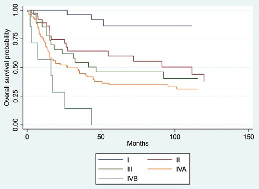
Figure 1. Kaplan-Meier curves for global survival by clinical stage.
Analysis by initial treatment showed significant differences in median overall survival (log-rank p adjusted for clinical stage = 0.001). Patients undergoing surgery alone reached 157.2 months, while those who received surgery with adjuvant treatment with radiotherapy or chemoradiotherapy presented 54.9 months. Patients who received induction chemotherapy followed by radiotherapy with or without adjuvant chemotherapy had a survival of 203.9 months, in contrast to those treated with definitive chemoradiotherapy, who achieved 31.4 months, and 15.2 months for patients treated with induction followed by surgery. Patients treated with exclusive radiotherapy or chemotherapy showed median survivals of 7.1 and 11.1 months, respectively. Figure 2 shows Kaplan-Meier curves for overall survival by treatment.
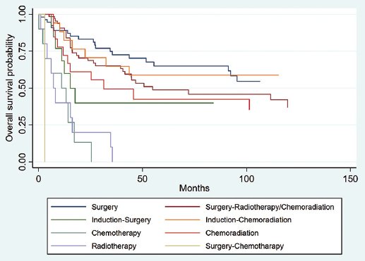
Figure 2. Overall survival by initial treatment sequence.
Univariate and multivariate analyses using the Cox model (Table 3) confirmed that age, functional status according to the ECOG scale, clinical stage at diagnosis, and type of treatment received were significant prognostic factors for overall survival, maintaining their relevance in both analyses.
Table 3. Univariate and multivariate analysis for global survival
| Variable | Univariate | Multivariate | ||||
|---|---|---|---|---|---|---|
| HR | 95% CI | p | HR | 95% CI | p | |
| Age | 1.02 | 1.01-1.04 | 0.001 | 1.02 | 1.01-1.04 | 0.003 |
| Gender | 1.21 | 0.82-1.79 | 0.330 | 0.87 | 0.58-1.31 | 0.509 |
| ECOG | 1.74 | 1.31-2.32 | 0.001 | 1.42 | 1.03-1.95 | 0.032 |
| Subsite | 1.04 | 0.92-1.16 | 0.531 | 0.99 | 0.92-1.06 | 0.787 |
| Stage | 1.60 | 1.32-1.93 | 0.001 | 1.44 | 1.15-1.79 | 0.001 |
| Treatment Surgery Surgery-Radiotherapy/Chemoradiation Induction-Surgery Induction-Chemoradiation Chemotherapy Chemoradiation Radiotherapy Surgery-Chemotherapy | 11.612.401.169.282.048.5568.43 | 10.93-2.781.01-5.670.51-2.624.10-20.981.00-4.153.94-18.587.87-594.61 | 10.0880.0460.7220.0010.0490.0010.001 | 11.151.310.773.751.234.0299.14 | 10.63-2.070.51-3.330.32-1.881.43-9.830.55-2.771.43-11.3010.06-976.54 | 10.6510.5700.5730.0070.6080.0080.001 |
|
ECOG: eastern cooperative oncology group; HR: hazard ratio; CI: confidence interval. |
||||||
Discussion
In our cohort, SCC represented 87.9% of cases, consistent with reported data. The proportion of advanced stages was 64% in Stages III, IVA, and IVB, which is slightly lower than the described 67-81%.10,11
The median overall survival in our cohort was 92.3 months, with marked variation by clinical stage. Our median survival for stage I was 111.6 months, while the median of 21.8 months in Stage IV reflects the limitations of treatment in advanced stages. This finding aligns with studies that also reported progressive decreases in survival with more advanced stages.12,13
Regarding treatment, we observed that patients treated solely with surgery had significantly greater survival compared to those who received definitive chemoradiotherapy or palliative treatments. This aligns with studies such as that by Wong et al., which highlight surgery as the intervention offering the best prognosis in early stages.14
Notably, the survival observed in patients treated with induction chemotherapy followed by radiotherapy (median of 203.9 months) was significantly higher than that reported in other studies, potentially influenced by factors such as patient selection with good functional status or previously mentioned factors.15,16 Although the use of induction chemotherapy is controversial, in our setting, it remains a therapeutic tool for locally advanced disease due to infrastructure capacity and delays in initiating radiotherapy treatment. However, the survival of patients treated with surgery after induction treatment was much lower, which has been previously reported;17 this may be explained by the risk of recurrence in the field of cancerization that is not easily delineated and, therefore, not completely resected.18,19
The main limitation of our study is its retrospective nature and the heterogeneity of the analyzed subgroups. However, it provides important real-world information regarding the treatment of patients with oral cavity cancer. The reported discrepancies highlight the need for additional studies to evaluate the implications of multiple contextual factors, early detection strategies, and therapeutic approaches that can be used in all regions.
Conclusions
Age, functional status, histology, and clinical stage at diagnosis are important prognostic factors in oral cavity cancer. Surgery remains the cornerstone of treatment for these patients. In advanced disease, induction chemotherapy followed by chemoradiotherapy is a treatment option.
Funding
The authors declare that they have not received funding.
Conflicts of interest
The authors declare no conflicts of interest.
Ethical considerations
Protection of human subjects and animals. The authors declare that no experiments on humans or animals were performed for this research.
Confidentiality, informed consent, and ethical approval. This study does not involve personal patient data, medical records, or biological samples, and does not require ethical approval. SAGER guidelines do not apply.
Declaration on the use of artificial intelligence. The authors declare that artificial intelligence was used in the writing of this manuscript. During the preparation of this work, the author(s) used OpenAI. (2024). ChatGPT retrieved from https://chat.openai.com/to correct spelling and grammar. After using this tool/service, the author(s) reviewed and edited the content as needed and take(s) full responsibility for the content of the published article.
